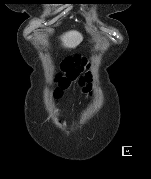
File:Calcified adrenal glands and Spigelian hernia (Radiopaedia 49741-54988 B 12).jpg
Jump to navigation
Jump to search
Size of this preview: 507 × 599 pixels. Other resolutions: 203 × 240 pixels | 512 × 605 pixels.
Original file (512 × 605 pixels, file size: 55 KB, MIME type: image/jpeg)
Summary:
| Description |
|
| Date | Published: 10th Dec 2016 |
| Source | https://radiopaedia.org/cases/calcified-adrenal-glands-and-spigelian-hernia |
| Author | Vikas Shah |
| Permission (Permission-reusing-text) |
http://creativecommons.org/licenses/by-nc-sa/3.0/ |
Licensing:
Attribution-NonCommercial-ShareAlike 3.0 Unported (CC BY-NC-SA 3.0)
File history
Click on a date/time to view the file as it appeared at that time.
| Date/Time | Thumbnail | Dimensions | User | Comment | |
|---|---|---|---|---|---|
| current | 03:37, 30 June 2021 | | 512 × 605 (55 KB) | Fæ (talk | contribs) | Radiopaedia project rID:49741 (batch #5750-262 B12) |
You cannot overwrite this file.
File usage
The following page uses this file:
.jpg&oldid=664790)