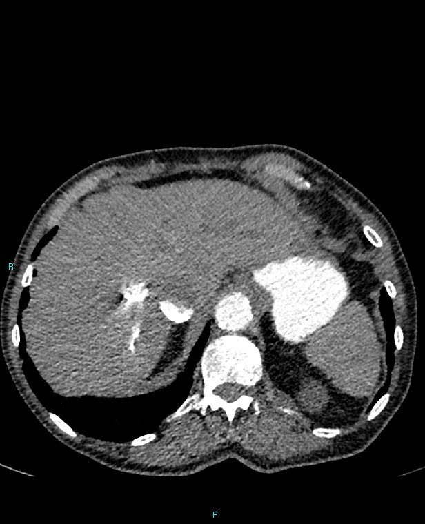
File:Calcified cerebral emboli from left ventricular thrombus (Radiopaedia 84420-99760 Axial C+ CTPA 78).jpg
Jump to navigation
Jump to search
Size of this preview: 486 × 600 pixels. Other resolutions: 194 × 240 pixels | 616 × 760 pixels.
Original file (616 × 760 pixels, file size: 86 KB, MIME type: image/jpeg)
Summary:
| Description |
|
| Date | Published: 24th Nov 2020 |
| Source | https://radiopaedia.org/cases/calcified-cerebral-emboli-from-left-ventricular-thrombus |
| Author | Tom Foster |
| Permission (Permission-reusing-text) |
http://creativecommons.org/licenses/by-nc-sa/3.0/ |
Licensing:
Attribution-NonCommercial-ShareAlike 3.0 Unported (CC BY-NC-SA 3.0)
File history
Click on a date/time to view the file as it appeared at that time.
| Date/Time | Thumbnail | Dimensions | User | Comment | |
|---|---|---|---|---|---|
| current | 05:45, 30 June 2021 | | 616 × 760 (86 KB) | Fæ (talk | contribs) | Radiopaedia project rID:84420 (batch #5763-78 A78) |
You cannot overwrite this file.
File usage
The following page uses this file:
.jpg&oldid=665623)