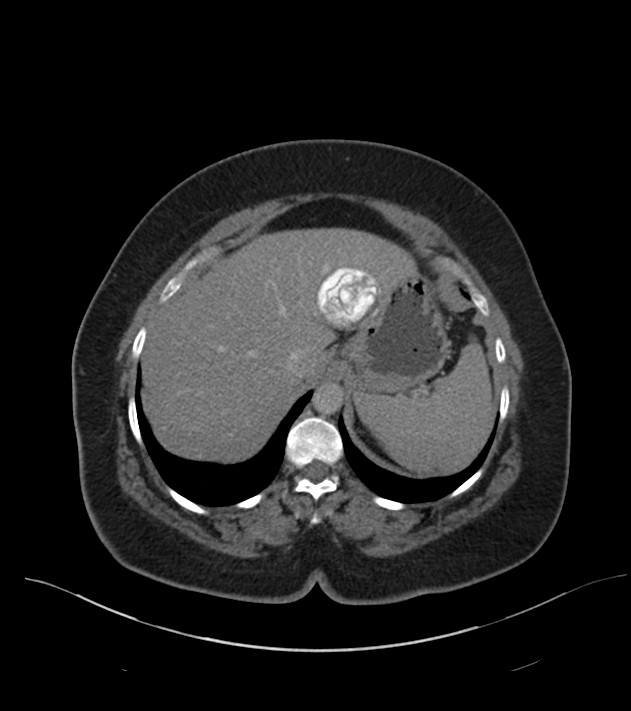
File:Calcified hepatic hydatid cyst (Radiopaedia 81127-94738 B 18).jpg
Jump to navigation
Jump to search
Size of this preview: 532 × 599 pixels. Other resolutions: 213 × 240 pixels | 631 × 711 pixels.
Original file (631 × 711 pixels, file size: 58 KB, MIME type: image/jpeg)
Summary:
| Description |
|
| Date | Published: 16th Aug 2020 |
| Source | https://radiopaedia.org/cases/calcified-hepatic-hydatid-cyst-2 |
| Author | Ahmed Abdelrahman |
| Permission (Permission-reusing-text) |
http://creativecommons.org/licenses/by-nc-sa/3.0/ |
Licensing:
Attribution-NonCommercial-ShareAlike 3.0 Unported (CC BY-NC-SA 3.0)
File history
Click on a date/time to view the file as it appeared at that time.
| Date/Time | Thumbnail | Dimensions | User | Comment | |
|---|---|---|---|---|---|
| current | 13:11, 30 June 2021 | | 631 × 711 (58 KB) | Fæ (talk | contribs) | Radiopaedia project rID:81127 (batch #5789-93 B18) |
You cannot overwrite this file.
File usage
The following page uses this file:
.jpg&oldid=668580)