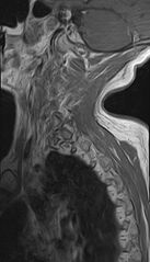
File:Calcified meningioma - cervical canal (Radiopaedia 70136-82468 Sagittal 2).jpg
Jump to navigation
Jump to search

Size of this preview: 343 × 599 pixels. Other resolutions: 137 × 240 pixels | 508 × 887 pixels.
Original file (508 × 887 pixels, file size: 164 KB, MIME type: image/jpeg)
Summary:
| Description |
|
| Date | Published: 2nd Nov 2019 |
| Source | https://radiopaedia.org/cases/calcified-meningioma-cervical-canal |
| Author | Dr Ammar Haouimi |
| Permission (Permission-reusing-text) |
http://creativecommons.org/licenses/by-nc-sa/3.0/ |
Licensing:
Attribution-NonCommercial-ShareAlike 3.0 Unported (CC BY-NC-SA 3.0)
File history
Click on a date/time to view the file as it appeared at that time.
| Date/Time | Thumbnail | Dimensions | User | Comment | |
|---|---|---|---|---|---|
| current | 17:37, 30 June 2021 | | 508 × 887 (164 KB) | Fæ (talk | contribs) | Radiopaedia project rID:70136 (batch #5816-53 E2) |
You cannot overwrite this file.
File usage
There are no pages that use this file.
.jpg&oldid=670331)