File:Calcium pyrophosphate dihydrate deposition disease (Radiopaedia 25455-25701 Axial bone window 26).png

From NC Commons
Jump to navigation Jump to search

Summary:

Description
  • Radiopaedia case ID: 25455
  • Study ID: 25701
  • Image ID: 4918711
  • Plane projection: Axial
  • Modality: CT
  • System: Trauma
  • Findings: There is calcification within the radiocapitellar, ulnotrochlear , and proximal radioulnar joints which parallels the articular surfaces to follow the contour of the hyaline cartilage. If one does not appreciate that this finding extends into the ulnotrochlear joint and proximal radioulnar joint, the chondrocalcinosis within the radiocapitellar joint could be mistaken for a radial head fracture in the setting of trauma. The absence of elevated fat pats mitigates this concern. In this case, the emergency department did mistakenly believe the patient had a radial head fracture based on the imaging. Remember that the hyaline cartilage of the radial head extends circumferentially around the radial head, so chondrocalcinosis would be expected to extend similarly to anywhere around the circumference of the radial head.
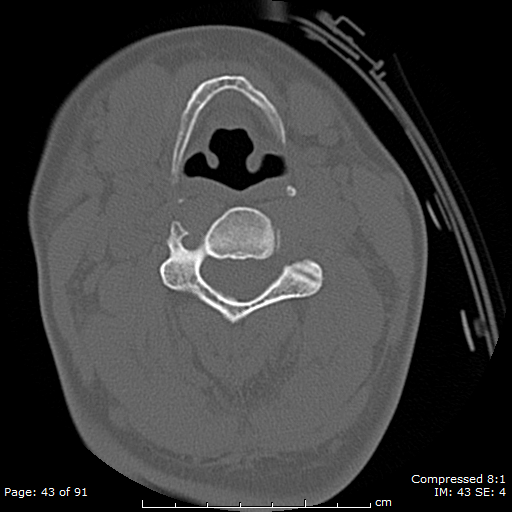
  • Study findings: Throughout the cervical spine, extensive calcification is observed within the disc spaces, as well as chondrocalcinosis within the uncovertebral joint spaces. Chondrocalcinosis is also seen within the synovial atlantoaxial joint spaces at the lateral masses, and around the dens in the atlantodental space. Calcification of the transverse ligament is also demonstrated. Chondrocalcinosis is observed within both of the atlantooccipital joints as well. There are multiple areas of calcification of the ligamentum flavum posteriorly within the neural canal, at the levels of  C3/4 and C5/6, with significant narrowing of the neural canal at C5/6. Thin but diffuse, faint areas of calcification of the thecal sac is noted.
  • Author location: San Jose, CA, United States
Date 26 Oct 2013
Source Calcium pyrophosphate dihydrate deposition disease
Author Matt Skalski
Permission
(Permission-reusing-text)
http://creativecommons.org/licenses/by-nc-sa/3.0/

Licensing:

Attribution-NonCommercial-ShareAlike 3.0 Unported (CC BY-NC-SA 3.0)

This file is ineligible for copyright and therefore in the public domain, because it is a technical image created as part of a standard medical diagnostic procedure. No creative element rising above the threshold of originality was involved in its production.

File history

Click on a date/time to view the file as it appeared at that time.

Date/TimeThumbnailDimensionsUserComment
current00:32, 1 July 2021Thumbnail for version as of 00:32, 1 July 2021512 × 512 (199 KB) (talk | contribs)Radiopaedia project rID:25455 (batch #5880-26 A26)

Metadata