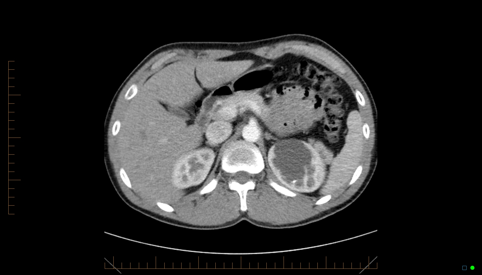
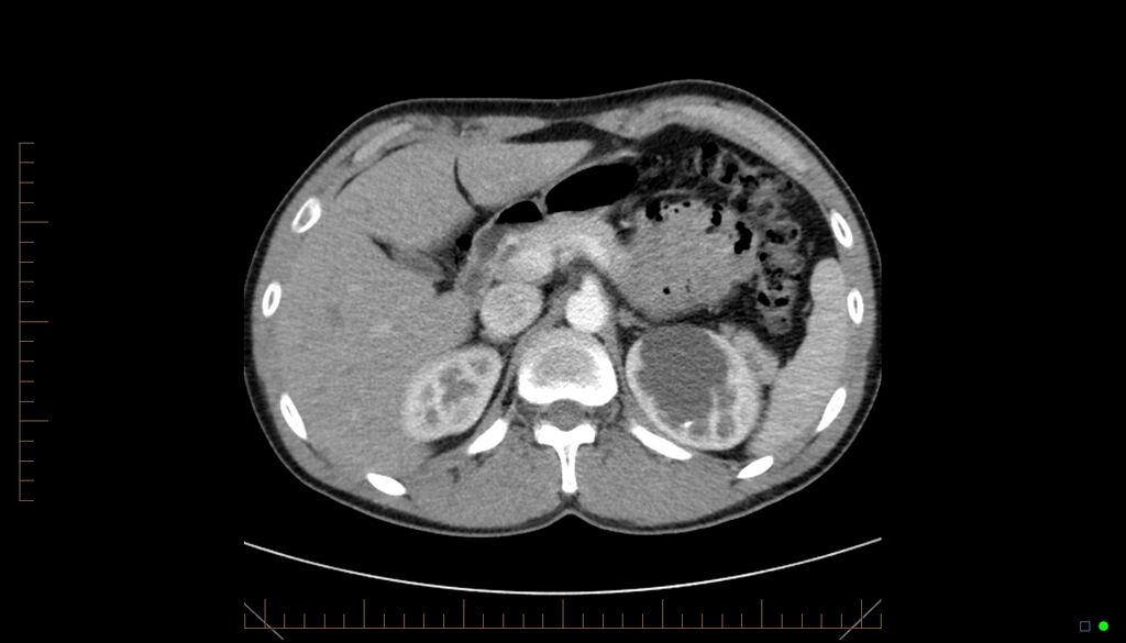
File:Calyceal diverticulum (Radiopaedia 35610-38112 Axial Multiphase 2).jpg
Jump to navigation
Jump to search
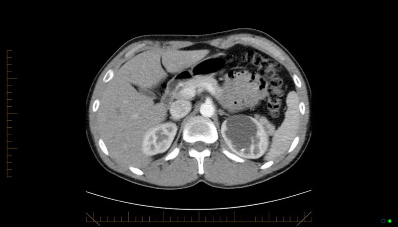
Size of this preview: 800 × 457 pixels. Other resolutions: 320 × 183 pixels | 640 × 365 pixels | 1,024 × 585 pixels | 1,625 × 928 pixels.
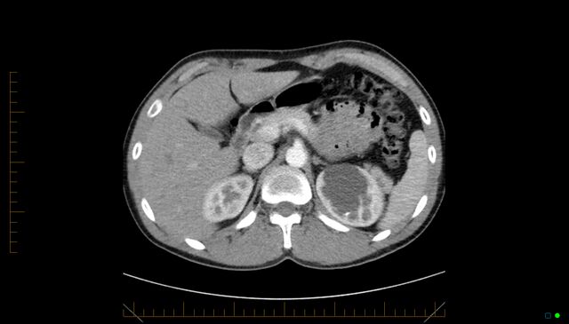
Original file (1,625 × 928 pixels, file size: 291 KB, MIME type: image/jpeg)
Summary:
| Description |
|
| Date | Published: 29th Apr 2015 |
| Source | https://radiopaedia.org/cases/calyceal-diverticulum-1 |
| Author | Wen Jak Ma |
| Permission (Permission-reusing-text) |
http://creativecommons.org/licenses/by-nc-sa/3.0/ |
Licensing:
Attribution-NonCommercial-ShareAlike 3.0 Unported (CC BY-NC-SA 3.0)
File history
Click on a date/time to view the file as it appeared at that time.
| Date/Time | Thumbnail | Dimensions | User | Comment | |
|---|---|---|---|---|---|
| current | 15:24, 1 July 2021 | | 1,625 × 928 (291 KB) | Fæ (talk | contribs) | Radiopaedia project rID:35610 (batch #5908-2 A2) |
You cannot overwrite this file.
File usage
There are no pages that use this file.
.jpg&oldid=675247)