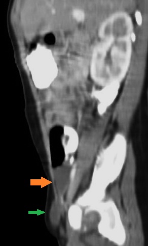
File:Canal of Nuck cyst (hydrocele) (Radiopaedia 41760-44715 A 1).jpg
Jump to navigation
Jump to search
No higher resolution available.
Canal_of_Nuck_cyst_(hydrocele)_(Radiopaedia_41760-44715_A_1).jpg (292 × 483 pixels, file size: 28 KB, MIME type: image/jpeg)
Summary:
| Description |
|
| Date | Published: 19th Dec 2015 |
| Source | https://radiopaedia.org/cases/canal-of-nuck-cyst-hydrocele-1 |
| Author | Rania Aly Zeitoun |
| Permission (Permission-reusing-text) |
http://creativecommons.org/licenses/by-nc-sa/3.0/ |
Licensing:
Attribution-NonCommercial-ShareAlike 3.0 Unported (CC BY-NC-SA 3.0)
File history
Click on a date/time to view the file as it appeared at that time.
| Date/Time | Thumbnail | Dimensions | User | Comment | |
|---|---|---|---|---|---|
| current | 17:23, 1 July 2021 | | 292 × 483 (28 KB) | Fæ (talk | contribs) | Radiopaedia project rID:41760 (batch #5925-1 A1) |
You cannot overwrite this file.
File usage
There are no pages that use this file.
_(Radiopaedia_41760-44715_A_1).jpg&oldid=676028)