File:Caput medusae sign - portal hypertension (Radiopaedia 64007-72759 A 92).jpg
Jump to navigation
Jump to search
No higher resolution available.
Caput_medusae_sign_-_portal_hypertension_(Radiopaedia_64007-72759_A_92).jpg (512 × 512 pixels, file size: 30 KB, MIME type: image/jpeg)
Summary:
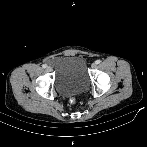
| Description |
|
| Date | Published: 30th Oct 2018 |
| Source | https://radiopaedia.org/cases/caput-medusae-sign-portal-hypertension |
| Author | Mohammad Taghi Niknejad |
| Permission (Permission-reusing-text) |
http://creativecommons.org/licenses/by-nc-sa/3.0/ |
Licensing:
Attribution-NonCommercial-ShareAlike 3.0 Unported (CC BY-NC-SA 3.0)
File history
Click on a date/time to view the file as it appeared at that time.
| Date/Time | Thumbnail | Dimensions | User | Comment | |
|---|---|---|---|---|---|
| current | 07:04, 2 July 2021 | | 512 × 512 (30 KB) | Fæ (talk | contribs) | Radiopaedia project rID:64007 (batch #5978-92 A92) |
You cannot overwrite this file.
File usage
The following page uses this file:
.jpg&oldid=681352)