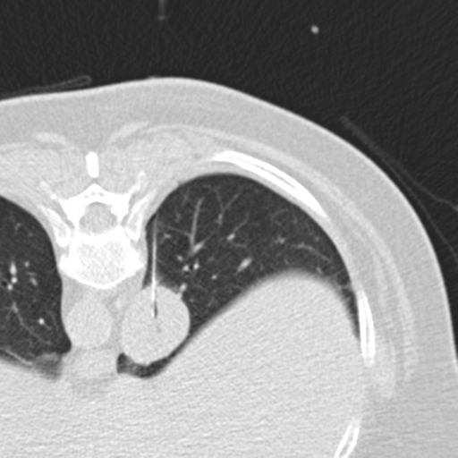
File:Carcinoid tumor - lung (Radiopaedia 44814-48644 Axial lung window 83).png
Jump to navigation
Jump to search
Carcinoid_tumor_-_lung_(Radiopaedia_44814-48644_Axial_lung_window_83).png (512 × 512 pixels, file size: 238 KB, MIME type: image/png)
Summary:
| Description |
|
| Date | Published: 5th May 2016 |
| Source | https://radiopaedia.org/cases/carcinoid-tumour-lung |
| Author | Bruno Di Muzio |
| Permission (Permission-reusing-text) |
http://creativecommons.org/licenses/by-nc-sa/3.0/ |
Licensing:
Attribution-NonCommercial-ShareAlike 3.0 Unported (CC BY-NC-SA 3.0)
File history
Click on a date/time to view the file as it appeared at that time.
| Date/Time | Thumbnail | Dimensions | User | Comment | |
|---|---|---|---|---|---|
| current | 11:35, 2 July 2021 |  | 512 × 512 (238 KB) | Fæ (talk | contribs) | Radiopaedia project rID:44814 (batch #5994-83 A83) |
You cannot overwrite this file.
File usage
The following page uses this file:
