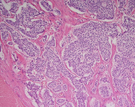
File:Carcinoid tumor of the ileum with metastasis to orbit (Radiopaedia 28165-28427 B 1).jpg

From NC Commons
Jump to navigation Jump to search

Carcinoid_tumor_of_the_ileum_with_metastasis_to_orbit_(Radiopaedia_28165-28427_B_1).jpg(514 × 410 pixels, file size: 102 KB, MIME type: image/jpeg)

Summary:

Description
  • Radiopaedia case ID: 28165
  • Image ID: 6220512
  • Plane projection: H&E
  • Study description: Histology (bowel)
  • Study findings: MACROSCOPIC DESCRIPTION: The specimen is a 300mm length of small bowel 20mm in diameter. Attached is mesentery along its entire length up to 50mm in thickness and 30mm in width. A polypoid brown tan nodule 35x25x15mm arises within the small bowel lumen. The nodule appears to push into but not through muscularis propria. The small bowel serosa is not affected and the lesion is 122mm from the closest resection margin. Multiple nodes are present in the small bowel mesentry. MICROSCOPIC DESCRIPTION: Sections show a neuroendocrine carcinoma extending into and focally through the muscularis propria. The tumor is composed of variably sized nests of mono-morphic polygonal cells with round nuclei and vesicular chromatin. There are up to 28 mitoses per 10 high power fields. There is no necrosis. Small nests are seen infiltrating through the muscularis propria and into adventitia with focal involvement of the serosal surface. Extensive lymphovascular invasion is identified. There is no perineural invasion. The mucosal margins are not involved by tumor. Metastatic tumor is present in 6 of 14 lymph nodes, including the apical lymph node. There are mutliple foci of extra-nodal extension. FINAL DIAGNOSIS: Well differentiated neuroendocrine carcinoma (AJCC pT4, pN2, M1 Stage IV 2). This tumor has been compared with the previous orbital mass and has the same morphological appearance. The findings are consistent with a primary small bowel neuroendocrine carcinoma metastastic to the orbit.
  • Modality: Pathology
  • System: Oncology
  • Findings: A well circumscribed right orbital mass demonstrating homogeneous contrast enhancement is seen. It displaces the medial rectus inferiorly and the superior rectus laterally. The optic nerve is displaced inferolaterally.
Date Published: 13th Mar 2014
Source https://radiopaedia.org/cases/carcinoid-tumour-of-the-ileum-with-metastasis-to-orbit
Author RMH Neuropathology
Permission
(Permission-reusing-text)
http://creativecommons.org/licenses/by-nc-sa/3.0/

Licensing:

Attribution-NonCommercial-ShareAlike 3.0 Unported (CC BY-NC-SA 3.0)

File history

Click on a date/time to view the file as it appeared at that time.

Date/TimeThumbnailDimensionsUserComment
current12:17, 2 July 2021Thumbnail for version as of 12:17, 2 July 2021514 × 410 (102 KB) (talk | contribs)Radiopaedia project rID:28165 (batch #5996-2 B1)

There are no pages that use this file.