File:Cardiac amyloidosis - ATTR wild-type (Radiopaedia 74412-85353 Look-Locker 22).jpg

From NC Commons
Jump to navigation Jump to search

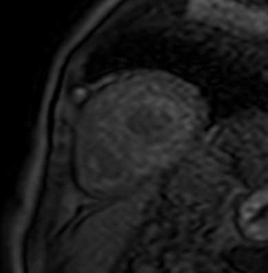
Cardiac_amyloidosis_-_ATTR_wild-type_(Radiopaedia_74412-85353_Look-Locker_22).jpg(389 × 397 pixels, file size: 24 KB, MIME type: image/jpeg)

Summary:

Description
  • Radiopaedia case ID: 74412
  • Image ID: 52063230
  • Image stack position: 22/45
  • Plane projection: Look-Locker
  • Study findings: Cine images, T2 black-blood and STIR imaging: Thickening of the interventricular septum (orange measurement >20mm) and the interatrial septum (blue measurement ≈ 8mm). T2 ratio was <1.9 – a T2 ratio of ≥2 is considered abnormal. Minimal pericardial effusion (blue arrowhead). Look-Locker: Images sorted by acquisition time. On close inspection, the myocardium passes through the zero-point before the blood pool. Usually, this is the other way around. Late gadolinium enhancement (LGE): It is conspicuous, that the blood pool has an even darker signal than the "nulled" myocardium not only on the inversion recovery gradient echo images but also on the PSIR image. This is due to high myocardial gadolinium uptake and fast blood washout referred to as abnormal blood-pool gadolinium kinetics 1. There is subendocardial late gadolinium enhancement in a non-coronary arterial distribution (red arrowheads) in midventricular and basal segments, and faint transmural enhancement of the septal and inferior basal segments (red arrows). T1 mapping native and postcontrast: Obviously increased native T1 here measured in the basal inferoseptal segment (z-score of 6.3). Native T1 measured in other segments ranged from 1170-1250 ms. Decreased postcontrast T1. Massively increased calculated extracellular volume (ECV) ≥60%.
  • Modality: Annotated image
  • System: Cardiac
  • Findings: Findings: Heart rate:  68 bpm, body surface area (BSA) 2.03 m² Hematocrit:  0.39 Image quality: no limitations Morphology and functional analysis: Global hypokinesia pronounced in the thickened basal inferoseptal and inferior segments. Mildly increased left atrium. Thickened atrial septum. Persistent foramen ovale (PFO). Aortic valve insufficiency with a diastolic jet in the left ventricular outflow tract (LVOT). No intracavitary thrombi found. Myocardial tissue properties Increased myocardial signal intensity in the apical segments probably due to stagnant blood. Difficulties in nulling the myocardium with the look locker sequence. PSIR and IR-GE sequences show a dark blood pool and a subendocardial late gadolinium enhancement in a non-coronary arterial distribution with a lesser extent in the apical segments, together with a transmural enhancement of the basal segments. T1 mapping native: 1170-1250 ms [950-1060ms*], extracellular volume (ECV): ≥60% *reference range based on local data No pericardial enhancement. Minimal pericardial effusion. Impression: Non-obstructive left ventricular hypertrophy with mild systolic dysfunction. Cardiac MRI findings are consistent with cardiac amyloidosis. The high values in extracellular volume (ECV) and the distinct thickening of the interventricular septum favor ATTR over AL cardiac amyloidosis.
Date Published: 24th Feb 2020
Source https://radiopaedia.org/cases/cardiac-amyloidosis-attr-wild-type-1
Author Joachim Feger
Permission
(Permission-reusing-text)
http://creativecommons.org/licenses/by-nc-sa/3.0/

Licensing:

Attribution-NonCommercial-ShareAlike 3.0 Unported (CC BY-NC-SA 3.0)

File history

Click on a date/time to view the file as it appeared at that time.

Date/TimeThumbnailDimensionsUserComment
current03:12, 3 July 2021Thumbnail for version as of 03:12, 3 July 2021389 × 397 (24 KB) (talk | contribs)Radiopaedia project rID:74412 (batch #6043-23 B22)

There are no pages that use this file.