File:Cardiac hemangioma (Radiopaedia 16971-16679 A 7).JPG

From NC Commons
Jump to navigation Jump to search

Original file(1,020 × 1,100 pixels, file size: 60 KB, MIME type: image/jpeg)

Summary:

Description
  • Radiopaedia case ID: 16971
  • Image ID: 1768503
  • Image stack position: 7/35
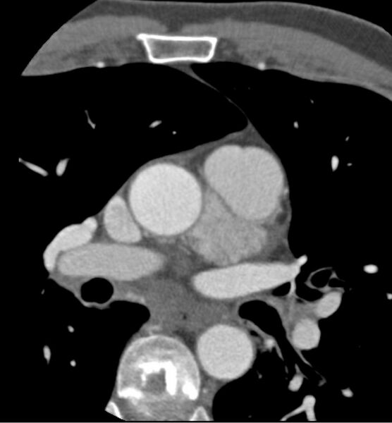
  • Plane projection: Axial
  • Aux modality: C+ arterial phase
  • Modality: CT
  • System: Cardiac
  • Findings: Preprocedurally, 5 mg of metoprolol was administered. The investigation itself provides the following features:Within the pericardium, a hypervascular structure is seen with a maximum diameter of 4.9 cm. Centrally, some hypodense calcifications are noted. Furthermore, the lesion is located between the pulmonary trunk, ascending aorta and the left pulmonary vein. It appears to drain via multiple venous structures that eventually meet in the coronary sinus. Obvious arterial blood supply cannot be observed. Also, no direct connection with the coronaries seems present. Some small branches of the right coronary artery might feed the lesion. However, no convincing connection is apparent. In contrast to the aorta and the coronaries, the lesion appears less dense. It does not seem to grow invasively. Moreover, the coronaries do not exhibit stenosis or vascular wall irregularities. N.B. the coronary arteries do not appear hyperplastic, as frequently seen in arteriovenous malformations or fistulas.
Date Published: 5th Mar 2012
Source https://radiopaedia.org/cases/cardiac-haemangioma
Author Radiology department - UMC Utrecht
Permission
(Permission-reusing-text)
http://creativecommons.org/licenses/by-nc-sa/3.0/

Licensing:

Attribution-NonCommercial-ShareAlike 3.0 Unported (CC BY-NC-SA 3.0)

File history

Click on a date/time to view the file as it appeared at that time.

Date/TimeThumbnailDimensionsUserComment
current04:32, 3 July 2021Thumbnail for version as of 04:32, 3 July 20211,020 × 1,100 (60 KB) (talk | contribs)Radiopaedia project rID:16971 (batch #6054-7 A7)

There are no pages that use this file.