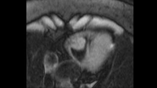
File:Cardiac hemangioma (Radiopaedia 31709-32631 F 1).jpg
Jump to navigation
Jump to search

Size of this preview: 800 × 450 pixels. Other resolutions: 320 × 180 pixels | 640 × 360 pixels | 1,280 × 720 pixels.
Original file (1,280 × 720 pixels, file size: 62 KB, MIME type: image/jpeg)
Summary:
| Description |
|
| Date | Published: 23rd Oct 2014 |
| Source | https://radiopaedia.org/cases/cardiac-haemangioma-1 |
| Author | Matt A. Morgan |
| Permission (Permission-reusing-text) |
http://creativecommons.org/licenses/by-nc-sa/3.0/ |
Licensing:
Attribution-NonCommercial-ShareAlike 3.0 Unported (CC BY-NC-SA 3.0)
| This file is ineligible for copyright and therefore in the public domain, because it is a technical image created as part of a standard medical diagnostic procedure. No creative element rising above the threshold of originality was involved in its production.
|
 |
File history
Click on a date/time to view the file as it appeared at that time.
| Date/Time | Thumbnail | Dimensions | User | Comment | |
|---|---|---|---|---|---|
| current | 05:00, 3 July 2021 | | 1,280 × 720 (62 KB) | Fæ (talk | contribs) | Radiopaedia project rID:31709 (batch #6055-114 F1) |
You cannot overwrite this file.
File usage
There are no pages that use this file.
.jpg&oldid=9754377)