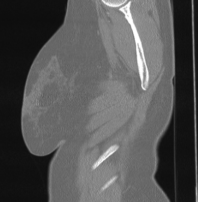
File:Cardiac sarcoidosis (Radiopaedia 74548-85534 Sagittal lung window 90).jpg

From NC Commons
Jump to navigation Jump to search

Cardiac_sarcoidosis_(Radiopaedia_74548-85534_Sagittal_lung_window_90).jpg(389 × 397 pixels, file size: 47 KB, MIME type: image/jpeg)

Summary:

Description
  • Radiopaedia case ID: 74548
  • Image ID: 52113252
  • Image stack position: 90/90
  • Plane projection: Sagittal
  • Aux modality: lung window
  • Study description: Thoracic CT
  • Study findings: Findings: Thyroid nodules (up 35 mm in size). Bilateral, symmetric hilar, right paratracheal and subcarinal lymph node enlargement, some lymph node calcifications. Pericardial effusion. Multiple small pulmonary (micro-)nodules with a perilymphatic distribution. Most are located along the fissures and the peribronchovascular interstitium and the subpleural surface. There is an upper and middle zone predominance. Spleen is not enlarged. Impression: CT-findings are consistent with pulmonary sarcoidosis. Multiple thyroid nodules, indicative of thyroid involvement. Pericardial effusion as an indicator of cardiac involvement.
  • Modality: CT
  • System: Cardiac
  • Findings: Findings: Heart rate:  58 bpm, BSA: 1,94 m² Image quality: no limitations Morphology and functional analysis: No regional left ventricular wall motion abnormalities. Visually normal atrial size. Persistent foramen ovale (PFO). Mild calcification of the anterior mitral leaflet. Mild mitral insufficiency. No intercavitary thrombi found. Myocardial tissue properties 1: No regional or global myocardial edema. Patchy intramyocardial and subepicardial late gadolinium enhancement (LGE) in the midventricular/apical inferior segments, best visible on the 2-chamber view (2ch). T2 mapping: 51 ms T1 mapping native: 990-1040 ms [950-1060ms*], extracellular volume (ECV):  < 30% *native T1 reference range based on local data Extensive, circular pericardial effusion. Impression: Patchy intramural and subepicardial late gadolinium enhancement visible in the apical/midventricular inferior segments and extensive pericardial effusion consistent with cardiac sarcoidosis. No signs of acute myocardial inflammation.
Date Published: 4th Mar 2020
Source https://radiopaedia.org/cases/cardiac-sarcoidosis-1
Author Joachim Feger
Permission
(Permission-reusing-text)
http://creativecommons.org/licenses/by-nc-sa/3.0/

Licensing:

Attribution-NonCommercial-ShareAlike 3.0 Unported (CC BY-NC-SA 3.0)

File history

Click on a date/time to view the file as it appeared at that time.

Date/TimeThumbnailDimensionsUserComment
current10:12, 3 July 2021Thumbnail for version as of 10:12, 3 July 2021389 × 397 (47 KB) (talk | contribs)Radiopaedia project rID:74548 (batch #6073-357 D90)