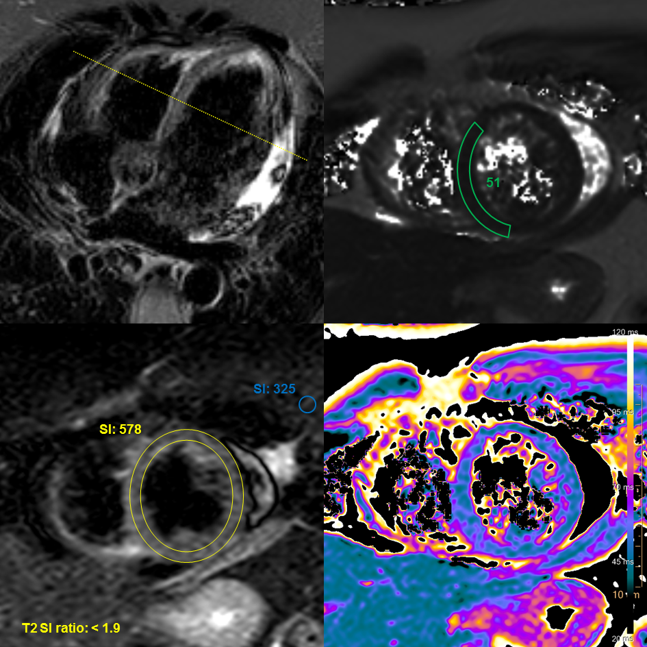
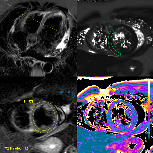
File:Cardiac sarcoidosis (Radiopaedia 74548-85645 STIR - T2 mapping 1).png

From NC Commons
Jump to navigation Jump to search

Original file(945 × 944 pixels, file size: 936 KB, MIME type: image/png)

Summary:

Description
  • Radiopaedia case ID: 74548
  • Image ID: 52107716
  • Plane projection: STIR - T2 mapping
  • Study findings: Short tau inversion recovery (STIR) imaging / T2 mapping: No focal myocardial edema can be seen. T2 signal intensity ratio between myocardium and skeletal muscle within the same image was calculated and was normal: <1.9 - a T2 SI ratio of ≥2 is considered abnormal, provided images were acquired with the MRI integrated body coil (Q-body) 2. T2 mapping:  51 ms (within normal range). T1 mapping native and postcontrast: native T1  ≈ 1025 ms, z-score of ≈ 0.8 (within normal limits) extracellular volume (ECV) was < 30% in all segments  ≈ 27.6 % (within normal limits) Late gadolinium enhancement (LGE): Patchy intramural/subepicardial late gadolinium enhancement can be seen in the inferior midventricular/apical segments on IR-GE images (red arrows) and reflects a granuloma or postinflammatory scar tissue.
  • Modality: Annotated image
  • System: Cardiac
  • Findings: Findings: Heart rate:  58 bpm, BSA: 1,94 m² Image quality: no limitations Morphology and functional analysis: No regional left ventricular wall motion abnormalities. Visually normal atrial size. Persistent foramen ovale (PFO). Mild calcification of the anterior mitral leaflet. Mild mitral insufficiency. No intercavitary thrombi found. Myocardial tissue properties 1: No regional or global myocardial edema. Patchy intramyocardial and subepicardial late gadolinium enhancement (LGE) in the midventricular/apical inferior segments, best visible on the 2-chamber view (2ch). T2 mapping: 51 ms T1 mapping native: 990-1040 ms [950-1060ms*], extracellular volume (ECV):  < 30% *native T1 reference range based on local data Extensive, circular pericardial effusion. Impression: Patchy intramural and subepicardial late gadolinium enhancement visible in the apical/midventricular inferior segments and extensive pericardial effusion consistent with cardiac sarcoidosis. No signs of acute myocardial inflammation.
Date Published: 4th Mar 2020
Source https://radiopaedia.org/cases/cardiac-sarcoidosis-1
Author Joachim Feger
Permission
(Permission-reusing-text)
http://creativecommons.org/licenses/by-nc-sa/3.0/

Licensing:

Attribution-NonCommercial-ShareAlike 3.0 Unported (CC BY-NC-SA 3.0)

File history

Click on a date/time to view the file as it appeared at that time.

Date/TimeThumbnailDimensionsUserComment
current09:19, 3 July 2021Thumbnail for version as of 09:19, 3 July 2021945 × 944 (936 KB) (talk | contribs)Radiopaedia project rID:74548 (batch #6073-1 A1)

There are no pages that use this file.

Metadata