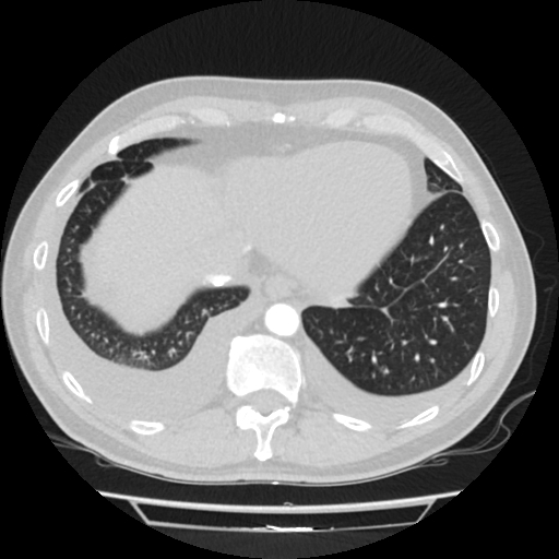
File:Cardiac tamponade (Radiopaedia 78607-91368 Axial lung window 76).jpg
Jump to navigation
Jump to search
Cardiac_tamponade_(Radiopaedia_78607-91368_Axial_lung_window_76).jpg (512 × 512 pixels, file size: 117 KB, MIME type: image/jpeg)
Summary:
| Description |
|
| Date | Published: 22nd Jun 2020 |
| Source | https://radiopaedia.org/cases/cardiac-tamponade-1 |
| Author | Rade Kovač |
| Permission (Permission-reusing-text) |
http://creativecommons.org/licenses/by-nc-sa/3.0/ |
Licensing:
Attribution-NonCommercial-ShareAlike 3.0 Unported (CC BY-NC-SA 3.0)
File history
Click on a date/time to view the file as it appeared at that time.
| Date/Time | Thumbnail | Dimensions | User | Comment | |
|---|---|---|---|---|---|
| current | 13:07, 3 July 2021 |  | 512 × 512 (117 KB) | Fæ (talk | contribs) | Radiopaedia project rID:78607 (batch #6077-398 C76) |
You cannot overwrite this file.
File usage
The following page uses this file:
