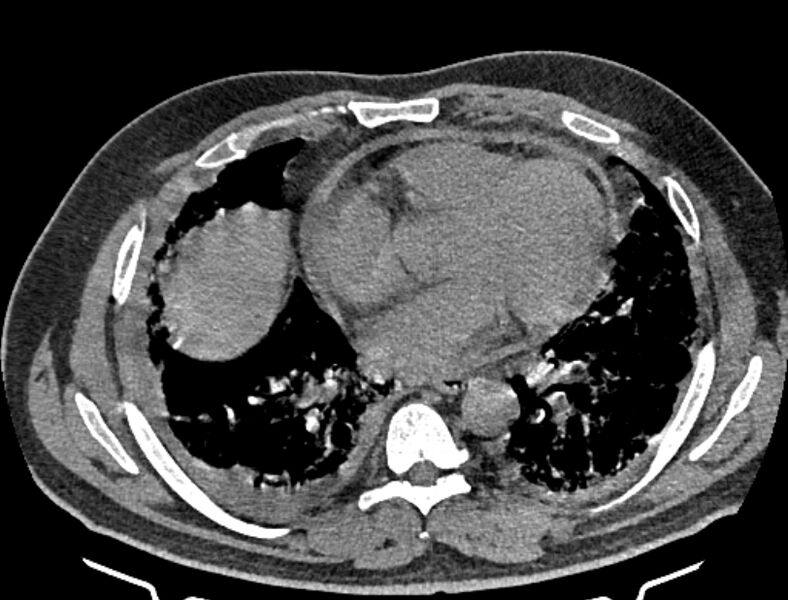
File:Cardiogenic pulmonary edema (Radiopaedia 68180-77678 Axial non-contrast 31).jpg
Jump to navigation
Jump to search

Size of this preview: 788 × 600 pixels. Other resolutions: 315 × 240 pixels | 631 × 480 pixels | 812 × 618 pixels.
Original file (812 × 618 pixels, file size: 246 KB, MIME type: image/jpeg)
Summary:
| Description |
|
| Date | Published: 6th Aug 2019 |
| Source | https://radiopaedia.org/cases/cardiogenic-pulmonary-oedema-2 |
| Author | Mostafa El-Feky |
| Permission (Permission-reusing-text) |
http://creativecommons.org/licenses/by-nc-sa/3.0/ |
Licensing:
Attribution-NonCommercial-ShareAlike 3.0 Unported (CC BY-NC-SA 3.0)
File history
Click on a date/time to view the file as it appeared at that time.
| Date/Time | Thumbnail | Dimensions | User | Comment | |
|---|---|---|---|---|---|
| current | 20:25, 3 July 2021 |  | 812 × 618 (246 KB) | Fæ (talk | contribs) | Radiopaedia project rID:68180 (batch #6087-31 A31) |
You cannot overwrite this file.
File usage
The following page uses this file: