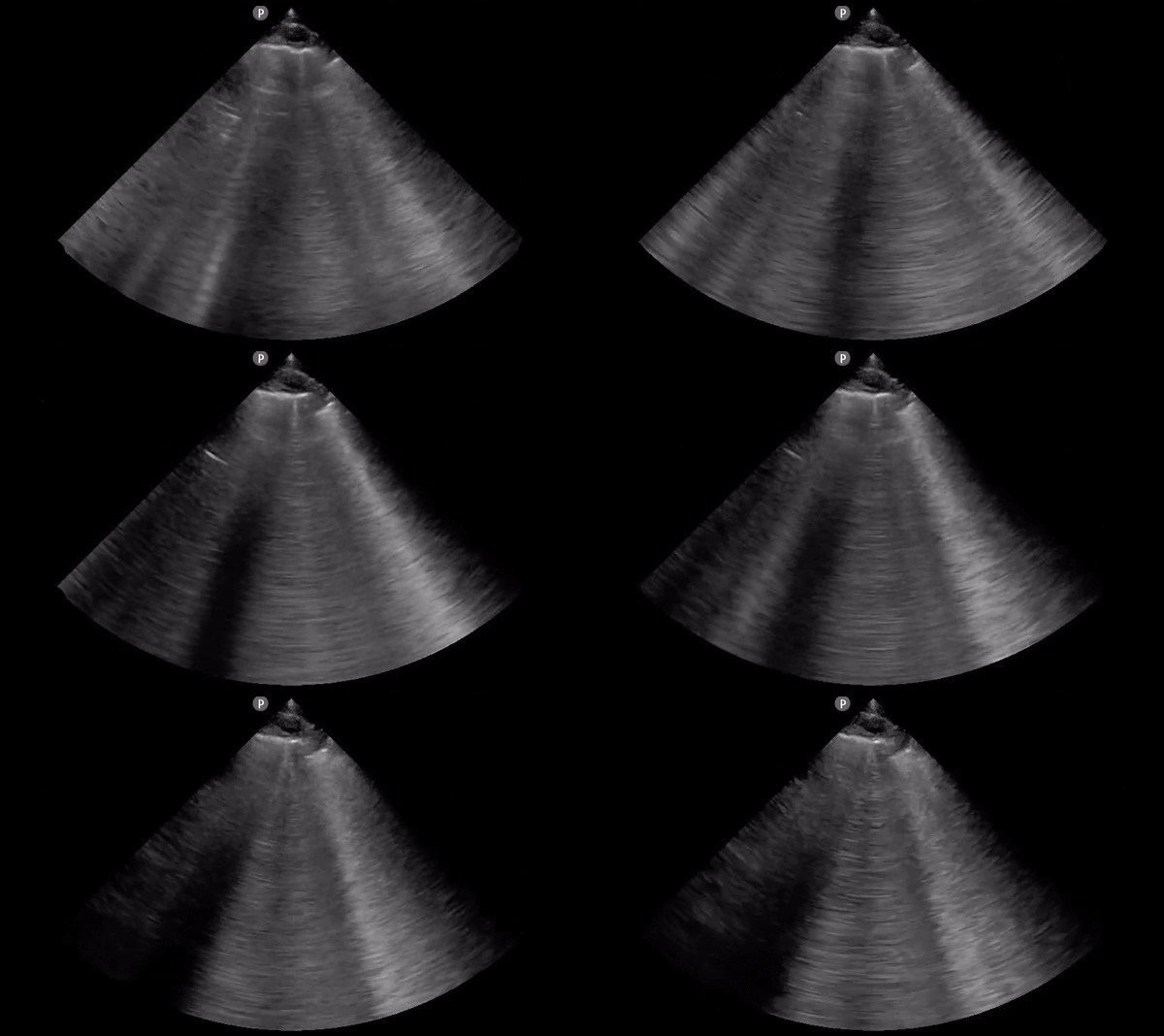
File:Cardiogenic pulmonary edema (ultrasound) (Radiopaedia 62735-71050 B 1).gif
Jump to navigation
Jump to search
Cardiogenic_pulmonary_edema_(ultrasound)_(Radiopaedia_62735-71050_B_1).gif (600 × 356 pixels, file size: 642 KB, MIME type: image/gif, looped, 15 frames, 1.7 s)
Summary:
| Description |
|
| Date | 29 Aug 2018 |
| Source | Cardiogenic pulmonary edema (ultrasound) |
| Author | David Carroll |
| Permission (Permission-reusing-text) |
http://creativecommons.org/licenses/by-nc-sa/3.0/ |
Licensing:
Attribution-NonCommercial-ShareAlike 3.0 Unported (CC BY-NC-SA 3.0)
| This file is ineligible for copyright and therefore in the public domain, because it is a technical image created as part of a standard medical diagnostic procedure. No creative element rising above the threshold of originality was involved in its production.
|
File history
Click on a date/time to view the file as it appeared at that time.
| Date/Time | Thumbnail | Dimensions | User | Comment | |
|---|---|---|---|---|---|
| current | 20:50, 3 July 2021 |  | 600 × 356 (642 KB) | Fæ (talk | contribs) | Radiopaedia project rID:62735 (batch #6090-2 B1) |
You cannot overwrite this file.
File usage
There are no pages that use this file.

