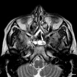
File:Caroticocavernous fistula (Radiopaedia 28599-28856 Axial T2 4).jpg
Jump to navigation
Jump to search
Caroticocavernous_fistula_(Radiopaedia_28599-28856_Axial_T2_4).jpg (256 × 256 pixels, file size: 15 KB, MIME type: image/jpeg)
Summary:
| Description |
|
| Date | Published: 7th Apr 2014 |
| Source | https://radiopaedia.org/cases/caroticocavernous-fistula-17 |
| Author | Abhinav Ranwaka |
| Permission (Permission-reusing-text) |
http://creativecommons.org/licenses/by-nc-sa/3.0/ |
Licensing:
Attribution-NonCommercial-ShareAlike 3.0 Unported (CC BY-NC-SA 3.0)
File history
Click on a date/time to view the file as it appeared at that time.
| Date/Time | Thumbnail | Dimensions | User | Comment | |
|---|---|---|---|---|---|
| current | 05:07, 4 July 2021 |  | 256 × 256 (15 KB) | Fæ (talk | contribs) | Radiopaedia project rID:28599 (batch #6126-17 B4) |
You cannot overwrite this file.
File usage
There are no pages that use this file.
