File:Carotid arterial dissection (Radiopaedia 30001-30548 B 110).jpg

From NC Commons
Jump to navigation Jump to search

Carotid_arterial_dissection_(Radiopaedia_30001-30548_B_110).jpg(512 × 512 pixels, file size: 16 KB, MIME type: image/jpeg)

Summary:

Description
  • Radiopaedia case ID: 30001
  • Image ID: 7530278
  • Image stack position: 110/130
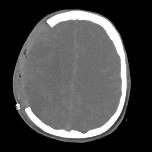
  • Plane projection: Axial
  • Aux modality: C+ arterial phase
  • Study description: CT at day 6
  • Study findings: Right hemicraniectomy with locules of gas and small pockets of hematoma laterally and inferiorly, seen within the overlying soft tissues. Extensive low attenuation within the territory of the right MCA, including the head of the right caudate nucleus is in keeping with extensive infarction. The right MCA is hyperdense in keeping with thrombosis. Approximately 2 cm distal to the bifurcation of the right common carotid, the enhancing lumen of contrast within the right internal carotid artery is significantly narrowed mildly irregular. The outer diameter of the ICA is, however, expanded, particularly at the base of skull, consistent with a dissection.
  • Modality: CT
  • System: Central Nervous System
  • Findings: Markedly hyperdense MCA is noted on the right associated with loss or grey - white matter differentiation in the temporal pole and insula cortex.
Date Published: 25th Jul 2016
Source https://radiopaedia.org/cases/carotid-arterial-dissection
Author Frank Gaillard
Permission
(Permission-reusing-text)
http://creativecommons.org/licenses/by-nc-sa/3.0/

Licensing:

Attribution-NonCommercial-ShareAlike 3.0 Unported (CC BY-NC-SA 3.0)

File history

Click on a date/time to view the file as it appeared at that time.

Date/TimeThumbnailDimensionsUserComment
current09:14, 4 July 2021Thumbnail for version as of 09:14, 4 July 2021512 × 512 (16 KB) (talk | contribs)Radiopaedia project rID:30001 (batch #6137-146 B110)

The following page uses this file: