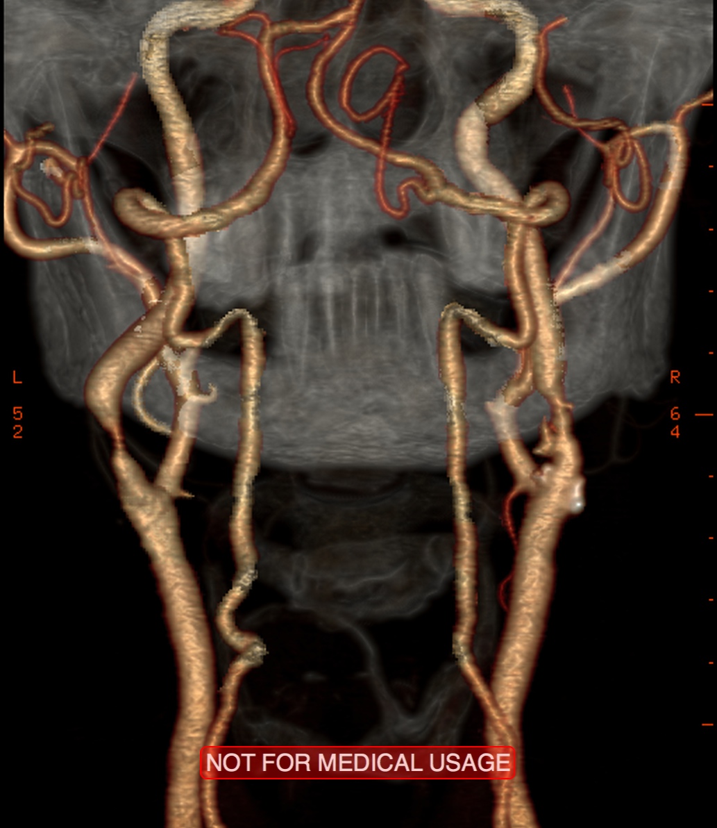
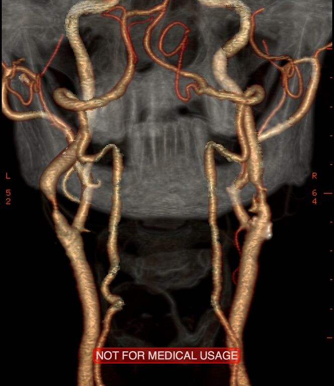
File:Carotid artery stenosis (Radiopaedia 28786-29086 A 8).jpg
Jump to navigation
Jump to search
Size of this preview: 519 × 599 pixels. Other resolutions: 208 × 240 pixels | 416 × 480 pixels | 665 × 768 pixels | 1,033 × 1,192 pixels.
Original file (1,033 × 1,192 pixels, file size: 259 KB, MIME type: image/jpeg)
Summary:
| Description |
|
| Date | Published: 12th Apr 2014 |
| Source | https://radiopaedia.org/cases/carotid-artery-stenosis-2 |
| Author | Bruno Di Muzio |
| Permission (Permission-reusing-text) |
http://creativecommons.org/licenses/by-nc-sa/3.0/ |
Licensing:
Attribution-NonCommercial-ShareAlike 3.0 Unported (CC BY-NC-SA 3.0)
File history
Click on a date/time to view the file as it appeared at that time.
| Date/Time | Thumbnail | Dimensions | User | Comment | |
|---|---|---|---|---|---|
| current | 12:55, 4 July 2021 | | 1,033 × 1,192 (259 KB) | Fæ (talk | contribs) | Radiopaedia project rID:28786 (batch #6149-8 A8) |
You cannot overwrite this file.
File usage
There are no pages that use this file.
.jpg&oldid=702149)