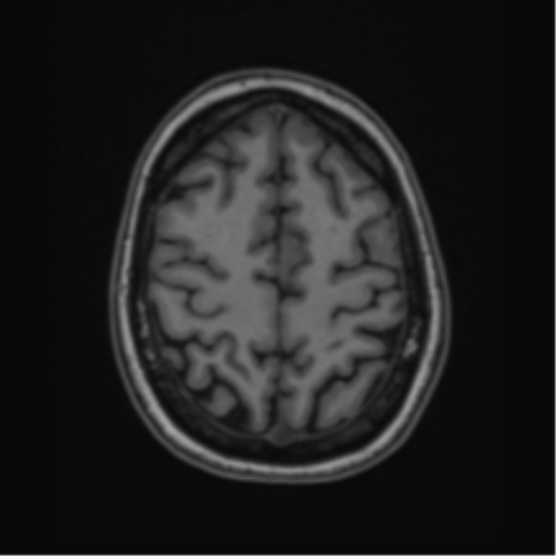
File:Cavernoma with bleed - midbrain (Radiopaedia 54546-60774 Axial T1 44).png
Jump to navigation
Jump to search
Cavernoma_with_bleed_-_midbrain_(Radiopaedia_54546-60774_Axial_T1_44).png (512 × 512 pixels, file size: 46 KB, MIME type: image/png)
Summary:
| Description |
|
| Date | Published: 23rd Jun 2018 |
| Source | https://radiopaedia.org/cases/cavernoma-with-bleed-midbrain |
| Author | Frank Gaillard |
| Permission (Permission-reusing-text) |
http://creativecommons.org/licenses/by-nc-sa/3.0/ |
Licensing:
Attribution-NonCommercial-ShareAlike 3.0 Unported (CC BY-NC-SA 3.0)
File history
Click on a date/time to view the file as it appeared at that time.
| Date/Time | Thumbnail | Dimensions | User | Comment | |
|---|---|---|---|---|---|
| current | 00:44, 8 July 2021 |  | 512 × 512 (46 KB) | Fæ (talk | contribs) | Radiopaedia project rID:54546 (batch #6345-106 C44) |
You cannot overwrite this file.
File usage
The following page uses this file:
