File:Cavernous hemangioma of the cerebellar falx (Radiopaedia 73025-83724 Sagittal FLAIR 134).jpg

From NC Commons
Jump to navigation Jump to search

Cavernous_hemangioma_of_the_cerebellar_falx_(Radiopaedia_73025-83724_Sagittal_FLAIR_134).jpg(512 × 512 pixels, file size: 99 KB, MIME type: image/jpeg)

Summary:

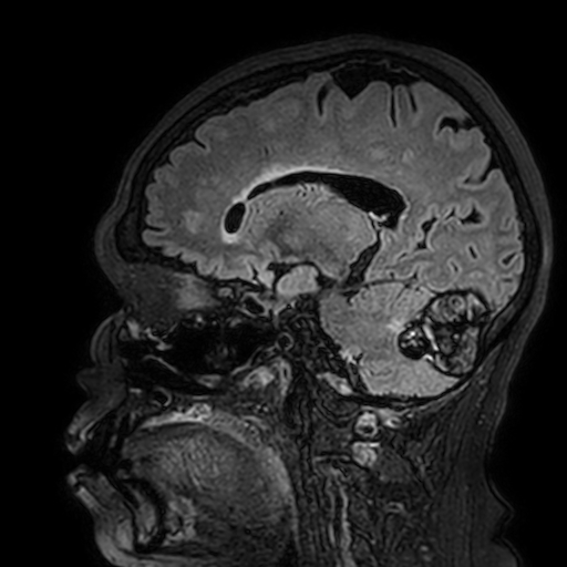
Description
  • Radiopaedia case ID: 73025
  • Image ID: 51853447
  • Image stack position: 134/215
  • Plane projection: Sagittal
  • Aux modality: FLAIR
  • Study description: MRI Brain with I/V Gadolinium
  • Study findings: A large (5.3 x 3.6 x 3.7 cm) well-defined midline-centered mass is identified posterosuperior to cerebellum, producing elevation of straight sinus, associated with sideways splitting and forward bulging of the falx cerebelli. After administration of I/V gadolinium, the split layers of falx cerebelli are well-identified wrapping around the lateral & anterior surfaces of the mass — confirming its location within the falx cerebelli. The mass itself and its walls show minimal or no enhancement. The mass is outlined by a well-defined lobulated outline containing multiple loculi. The loculi demonstrate mixed iso-, hyper- & hypo-intense as well as a signal-void appearance on T1, T2 & FLAIR images, followed by blooming on BOLD images, thus representing hemoglobin derivatives and blood-blood levels. The mass compresses upon cerebellar vermis & both hemispheres as well as fourth ventricle. An ill-defined thin hyperintense zone of perilesional edema is seen along the compressed posterior surface of cerebellum. Rest of the ventricles and extraventricular CSF spaces appear widened. However, no diffuse hyperintensity is noted in paraventricular white matter.
  • Modality: MRI
  • System: Vascular
  • Findings: A large (5.3 x 3.6 x 3.7 cm) well-defined midline-centered mass is identified posterosuperior to cerebellum, producing apparent splitting of the falx cerebelli. It has a sharp lobulated outline, containing multiple loculi of mixed hyper & hypodense appearance. The mass compresses upon cerebellar vermis & both hemispheres as well as fourth ventricle. Rest of the ventricles and extraventricular CSF spaces appear widened.
Date Published: 1st Jan 2020
Source https://radiopaedia.org/cases/cavernous-haemangioma-of-the-cerebellar-falx
Author Muhammad Shoyab
Permission
(Permission-reusing-text)
http://creativecommons.org/licenses/by-nc-sa/3.0/

Licensing:

Attribution-NonCommercial-ShareAlike 3.0 Unported (CC BY-NC-SA 3.0)

File history

Click on a date/time to view the file as it appeared at that time.

Date/TimeThumbnailDimensionsUserComment
current09:10, 8 July 2021Thumbnail for version as of 09:10, 8 July 2021512 × 512 (99 KB) (talk | contribs)Radiopaedia project rID:73025 (batch #6356-422 E134)