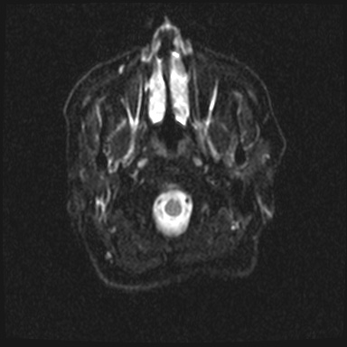
File:Cavernous sinus meningioma (Radiopaedia 63682-72367 DWI 2).jpg
Jump to navigation
Jump to search
Cavernous_sinus_meningioma_(Radiopaedia_63682-72367_DWI_2).jpg (384 × 384 pixels, file size: 38 KB, MIME type: image/jpeg)
Summary:
| Description |
|
| Date | Published: 13th Oct 2018 |
| Source | https://radiopaedia.org/cases/cavernous-sinus-meningioma-5 |
| Author | Eid Kakish |
| Permission (Permission-reusing-text) |
http://creativecommons.org/licenses/by-nc-sa/3.0/ |
Licensing:
Attribution-NonCommercial-ShareAlike 3.0 Unported (CC BY-NC-SA 3.0)
File history
Click on a date/time to view the file as it appeared at that time.
| Date/Time | Thumbnail | Dimensions | User | Comment | |
|---|---|---|---|---|---|
| current | 19:05, 8 July 2021 |  | 384 × 384 (38 KB) | Fæ (talk | contribs) | Radiopaedia project rID:63682 (batch #6371-102 F2) |
You cannot overwrite this file.
File usage
The following page uses this file:
