File:Cavernous transformation of the portal vein (Radiopaedia 53691-59751 B 16).jpg

From NC Commons
Jump to navigation Jump to search

Cavernous_transformation_of_the_portal_vein_(Radiopaedia_53691-59751_B_16).jpg(512 × 433 pixels, file size: 106 KB, MIME type: image/jpeg)

Summary:

Description
  • Radiopaedia case ID: 53691
  • Image ID: 30662872
  • Image stack position: 16/56
  • Plane projection: Coronal
  • Aux modality: C+ portal venous phase
  • Modality: CT
  • System: Hepatobiliary
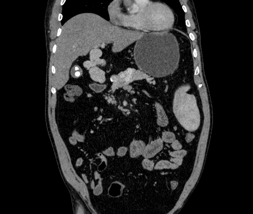
  • Findings: Replacement of the normal single channel portal vein with numerous tortuous enhancing collateral vessels are seen in the peripancreatic region and porta hepatis indicative of cavernous transformation of the portal vein. Draining into the collateral vessels is the dilated splenic vein. The superior mesenteric vein and its tributaries are likewise dilated and appear to drain into the afore mentioned collaterals. The right and left intrahepatic branches of the portal vein are attenuated. The liver shows mild atrophic left lateral segments (segment II and III) with hypertrophy of the caudate lobe and segment IV,  the liver shows fat density. Wandering spleen is mildly enlarged and seen at the left lumbar region. Calculous cholecystitis. Inflammatory bowel disease in the form of fat submucosal deposition seen in the descending colon, sigmoid colon and the rectum (fat halo sign), with diffuse loss of the colonic haustrations.
Date Published: 1st Jun 2017
Source https://radiopaedia.org/cases/cavernous-transformation-of-the-portal-vein-11
Author Mohammad Farghali Ali Tosson
Permission
(Permission-reusing-text)
http://creativecommons.org/licenses/by-nc-sa/3.0/

Licensing:

Attribution-NonCommercial-ShareAlike 3.0 Unported (CC BY-NC-SA 3.0)

File history

Click on a date/time to view the file as it appeared at that time.

Date/TimeThumbnailDimensionsUserComment
current23:13, 8 July 2021Thumbnail for version as of 23:13, 8 July 2021512 × 433 (106 KB) (talk | contribs)Radiopaedia project rID:53691 (batch #6380-80 B16)