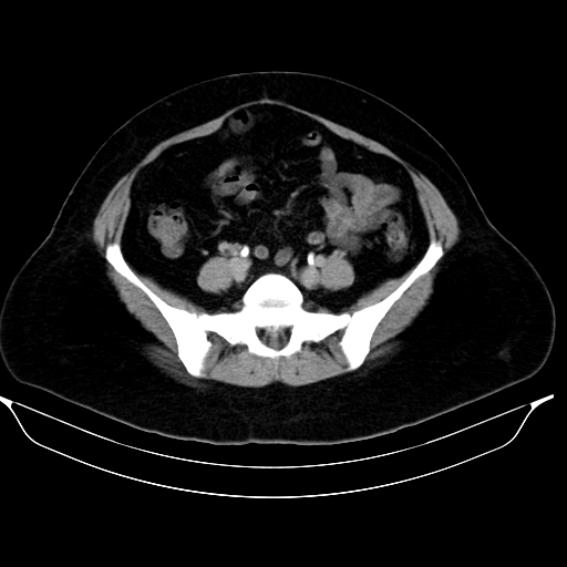
File:Cavernous transformation of the portal vein (Radiopaedia 58933-66189 Axial C+ delayed 29).jpg
Jump to navigation
Jump to search
Cavernous_transformation_of_the_portal_vein_(Radiopaedia_58933-66189_Axial_C+_delayed_29).jpg (512 × 512 pixels, file size: 96 KB, MIME type: image/jpeg)
Summary:
| Description |
|
| Date | Published: 13th Mar 2018 |
| Source | https://radiopaedia.org/cases/cavernous-transformation-of-the-portal-vein-13 |
| Author | Amr Farouk |
| Permission (Permission-reusing-text) |
http://creativecommons.org/licenses/by-nc-sa/3.0/ |
Licensing:
Attribution-NonCommercial-ShareAlike 3.0 Unported (CC BY-NC-SA 3.0)
File history
Click on a date/time to view the file as it appeared at that time.
| Date/Time | Thumbnail | Dimensions | User | Comment | |
|---|---|---|---|---|---|
| current | 00:48, 9 July 2021 |  | 512 × 512 (96 KB) | Fæ (talk | contribs) | Radiopaedia project rID:58933 (batch #6385-139 D29) |
You cannot overwrite this file.
File usage
There are no pages that use this file.
