File:Cavitating pneumonia (Radiopaedia 18359).jpg
Jump to navigation
Jump to search

Size of this preview: 493 × 599 pixels. Other resolutions: 197 × 240 pixels | 395 × 480 pixels | 632 × 768 pixels | 842 × 1,024 pixels | 1,760 × 2,140 pixels.
Original file (1,760 × 2,140 pixels, file size: 181 KB, MIME type: image/jpeg)
Summary:
- Radiopaedia case ID: 18359
- Image ID: 2081598
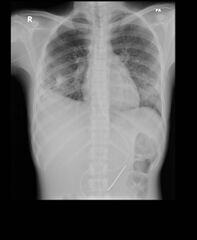
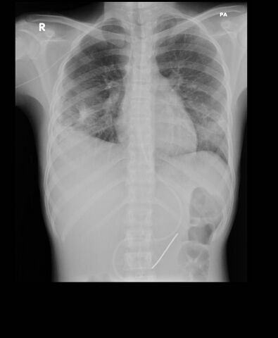
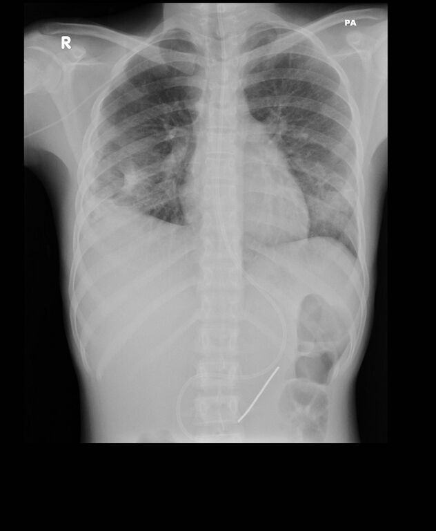
- Study findings: Bilateral cavitating lesions are present in the lower zones consistent with cavitating pneumonia.Pleural thickening is noted on the right.
- Modality: X-ray
- System: Chest
- Findings: Bilateral cavitating lesions are present in the lower zones consistent with cavitating pneumonia. Pleural thickening is noted on the right.
- Published: 3rd Jul 2012
- Source: https://radiopaedia.org/cases/cavitating-pneumonia-1
- Author: Townsville radiology training
- Permission: http://creativecommons.org/licenses/by-nc-sa/3.0/
Licensing:
Attribution-NonCommercial-ShareAlike 3.0 Unported (CC BY-NC-SA 3.0)
File history
Click on a date/time to view the file as it appeared at that time.
| Date/Time | Thumbnail | Dimensions | User | Comment | |
|---|---|---|---|---|---|
| current | 12:26, 20 March 2021 | | 1,760 × 2,140 (181 KB) | Fæ (talk | contribs) | Radiopaedia project rID:18359 (batch #6193) |
You cannot overwrite this file.
File usage
There are no pages that use this file.
.jpg&oldid=8859286)