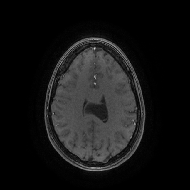
File:Cavum velum interpositum cyst (Radiopaedia 21593-21548 MRA 11).jpg
Jump to navigation
Jump to search
Size of this preview: 600 × 600 pixels. Other resolutions: 240 × 240 pixels | 480 × 480 pixels | 647 × 647 pixels.
Original file (647 × 647 pixels, file size: 57 KB, MIME type: image/jpeg)
Summary:
| Description |
|
| Date | Published: 2nd Feb 2013 |
| Source | https://radiopaedia.org/cases/cavum-velum-interpositum-cyst-1 |
| Author | Alexandra Stanislavsky |
| Permission (Permission-reusing-text) |
http://creativecommons.org/licenses/by-nc-sa/3.0/ |
Licensing:
Attribution-NonCommercial-ShareAlike 3.0 Unported (CC BY-NC-SA 3.0)
File history
Click on a date/time to view the file as it appeared at that time.
| Date/Time | Thumbnail | Dimensions | User | Comment | |
|---|---|---|---|---|---|
| current | 22:09, 9 July 2021 | | 647 × 647 (57 KB) | Fæ (talk | contribs) | Radiopaedia project rID:21593 (batch #6450-103 F11) |
You cannot overwrite this file.
File usage
The following page uses this file:
.jpg&oldid=736222)