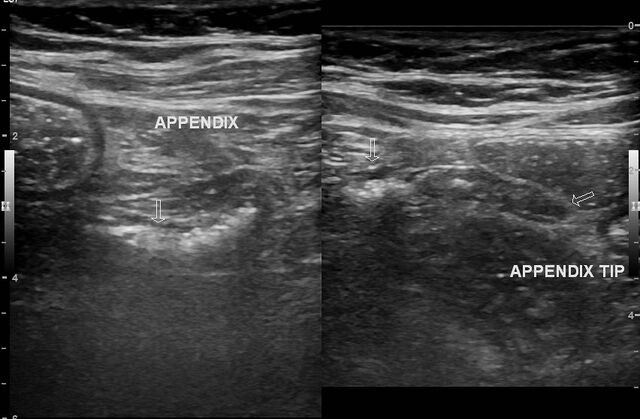
File:Cecal diverticulitis (Radiopaedia 88377).jpg
Jump to navigation
Jump to search

Size of this preview: 800 × 524 pixels. Other resolutions: 320 × 210 pixels | 640 × 419 pixels | 852 × 558 pixels.
Original file (852 × 558 pixels, file size: 109 KB, MIME type: image/jpeg)
Summary:
| Description |
|
| Date | Published: 6th Apr 2021 |
| Source | https://radiopaedia.org/cases/caecal-diverticulitis-3 |
| Author | Maulik S Patel |
| Permission (Permission-reusing-text) |
http://creativecommons.org/licenses/by-nc-sa/3.0/ |
Licensing:
Attribution-NonCommercial-ShareAlike 3.0 Unported (CC BY-NC-SA 3.0)
File history
Click on a date/time to view the file as it appeared at that time.
| Date/Time | Thumbnail | Dimensions | User | Comment | |
|---|---|---|---|---|---|
| current | 17:35, 28 June 2021 | | 852 × 558 (109 KB) | Fæ (talk | contribs) | Radiopaedia project rID:88377 (batch #5539) |
You cannot overwrite this file.
File usage
The following page uses this file:
.jpg&oldid=8853455)