File:Cecal mass causing appendicitis (Radiopaedia 59207-66532 K 34).jpg

From NC Commons
Jump to navigation Jump to search

Cecal_mass_causing_appendicitis_(Radiopaedia_59207-66532_K_34).jpg(320 × 260 pixels, file size: 15 KB, MIME type: image/jpeg)

Summary:

Description
  • Radiopaedia case ID: 59207
  • Image ID: 37228655
  • Image stack position: 34/80
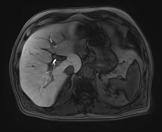
  • Plane projection: Axial
  • Aux modality: T1 C+ 20 minutes
  • Study description: Primovist MRI liver
  • Study findings: Segment 7/8 lesion identified on previous CT demonstrates intense high T2 signal, high signal on high b-value DWI and progressive peripheral nodular enhancement from the arterial to venous phases. There is no uptake of Primovist in this lesion. A further small arterially hyperenhancing lesion is demonstrated in segment 3, with persisting enhancement into the venous phases and no uptake of Primovist. These are both likely hemangiomas.
  • Modality: MRI
  • System: Gastrointestinal
  • Findings: The appendix is dilated and thick walled, with surrounding stranding in keeping with acute appendicitis. Moderate surrounding inflammatory change, with no extraluminal gas or collection. At the base of the appendix in the cecum, there is a hyperdense mass. Pericaecal fat stranding is also demonstrated. Enlarged ileocecal lymph nodes are present. In the right liver there is a hypodense lesion. Conclusion: Suspected cecal cancer obstructing the appendiceal orifice, with acute appendicitis. Right liver hypodense lesion in this context is suspicious for a metastasis. MRI liver is suggested for further evaluation and to identify any other metastases.
Date Published: 27th Mar 2018
Source https://radiopaedia.org/cases/caecal-mass-causing-appendicitis
Author Wayland Wang
Permission
(Permission-reusing-text)
http://creativecommons.org/licenses/by-nc-sa/3.0/

Licensing:

Attribution-NonCommercial-ShareAlike 3.0 Unported (CC BY-NC-SA 3.0)

File history

Click on a date/time to view the file as it appeared at that time.

Date/TimeThumbnailDimensionsUserComment
current20:44, 28 June 2021Thumbnail for version as of 20:44, 28 June 2021320 × 260 (15 KB) (talk | contribs)Radiopaedia project rID:59207 (batch #5543-639 K34)