File:Cecal volvulus (Radiopaedia 51046-56620 A 37).png

From NC Commons
Jump to navigation Jump to search

Cecal_volvulus_(Radiopaedia_51046-56620_A_37).png(512 × 512 pixels, file size: 56 KB, MIME type: image/png)

Summary:

Description
  • Radiopaedia case ID: 51046
  • Image ID: 28135414
  • Image stack position: 37/87
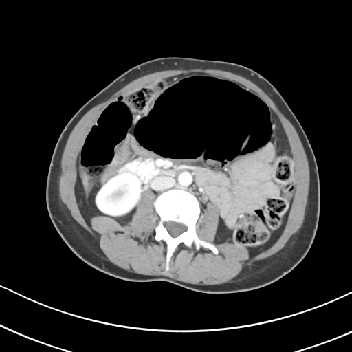
  • Plane projection: Axial
  • Aux modality: C+ portal venous phase
  • Modality: CT
  • System: Gastrointestinal
  • Findings: A large dilated loop of colon is noted in the central abdomen appearing to arise from the right iliac fossa measuring up to 9 cm. A swirl of mesenteric vessels is noted in the right iliac fossa leading into the dilated bowel. There is a blind ending tubular structure arising from the tip of the distended bowel, compatible with an appendix arising from the cecal tip. A moderate volume of peritoneal free fluid is present. No free gas. The remainder of the colon is unremarkable. No dilated small bowel loops. The liver, spleen, pancreas, adrenals and kidneys are unremarkable. Stomach is distended with oral contrast. Imaged lung bases are clear. No suspicious osseous lesions. Conclusion: Findings in keeping with cecal volvulus, dilated up to 9 cm, with peritoneal free fluid but no free gas.
Date Published: 27th Mar 2018
Source https://radiopaedia.org/cases/caecal-volvulus-15
Author Wayland Wang
Permission
(Permission-reusing-text)
http://creativecommons.org/licenses/by-nc-sa/3.0/

Licensing:

Attribution-NonCommercial-ShareAlike 3.0 Unported (CC BY-NC-SA 3.0)

File history

Click on a date/time to view the file as it appeared at that time.

Date/TimeThumbnailDimensionsUserComment
current22:38, 28 June 2021Thumbnail for version as of 22:38, 28 June 2021512 × 512 (56 KB) (talk | contribs)Radiopaedia project rID:51046 (batch #5548-37 A37)

The following page uses this file:

Metadata