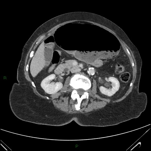
File:Cecal volvulus (Radiopaedia 78953-91866 A 100).jpg
Jump to navigation
Jump to search
Cecal_volvulus_(Radiopaedia_78953-91866_A_100).jpg (512 × 512 pixels, file size: 47 KB, MIME type: image/jpeg)
Summary:
| Description |
|
| Date | Published: 18th Jun 2020 |
| Source | https://radiopaedia.org/cases/caecal-volvulus-28 |
| Author | Ian Bickle |
| Permission (Permission-reusing-text) |
http://creativecommons.org/licenses/by-nc-sa/3.0/ |
Licensing:
Attribution-NonCommercial-ShareAlike 3.0 Unported (CC BY-NC-SA 3.0)
File history
Click on a date/time to view the file as it appeared at that time.
| Date/Time | Thumbnail | Dimensions | User | Comment | |
|---|---|---|---|---|---|
| current | 00:32, 29 June 2021 |  | 512 × 512 (47 KB) | Fæ (talk | contribs) | Radiopaedia project rID:78953 (batch #5552-100 A100) |
You cannot overwrite this file.
File usage
The following page uses this file:
