File:Cecal volvulus (Radiopaedia 86741-102900 B 49).jpg

From NC Commons
Jump to navigation Jump to search

Cecal_volvulus_(Radiopaedia_86741-102900_B_49).jpg(224 × 252 pixels, file size: 15 KB, MIME type: image/jpeg)

Summary:

Description
  • Radiopaedia case ID: 86741
  • Image ID: 54371324
  • Image stack position: 49/77
  • Plane projection: Coronal
  • Aux modality: C+ portal venous phase
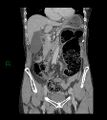
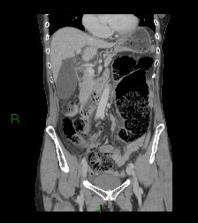
  • Study findings: Distended cecum measuring up to 10.5 cm in diameter. Terminal ileum and ileocecal valve are relatively superior in the abdomen. Sharp transition point of the cecum inferiorly, with apparent 'whirlpool' appearance of the mesentery at the transition point. The large bowel distal to the transition point is entirely collapsed. Fecalised material in the distal ileum. Small volume free abdominal and pelvic fluid. No discrete collection. No free gas.Gallstone in an otherwise unremarkable gallbladder.Normal liver, adrenals, kidneys, pancreas and spleen.Severe T9 and moderate T12 osteoporotic wedge fractures. No concerning bony abnormality.Opinion:Appearances in keeping with cecal volvulus, with cecum measuring up to 10.5 cm in diameter. No perforation.
  • Modality: CT
  • System: Gastrointestinal
  • Findings: Distended loop of large bowel measuring up to 11 cm projected over the left side of the abdomen. No free intraperitoneal gas.
Date Published: 11th Feb 2021
Source https://radiopaedia.org/cases/caecal-volvulus-26
Author Matthew Tse
Permission
(Permission-reusing-text)
http://creativecommons.org/licenses/by-nc-sa/3.0/

Licensing:

Attribution-NonCommercial-ShareAlike 3.0 Unported (CC BY-NC-SA 3.0)

File history

Click on a date/time to view the file as it appeared at that time.

Date/TimeThumbnailDimensionsUserComment
current02:26, 29 June 2021Thumbnail for version as of 02:26, 29 June 2021224 × 252 (15 KB) (talk | contribs)Radiopaedia project rID:86741 (batch #5558-200 B49)

The following page uses this file:

Metadata