File:Cecal volvulus (Radiopaedia 89212-106088 B 53).jpg

From NC Commons
Jump to navigation Jump to search

Cecal_volvulus_(Radiopaedia_89212-106088_B_53).jpg(512 × 512 pixels, file size: 74 KB, MIME type: image/jpeg)

Summary:

Description
  • Radiopaedia case ID: 89212
  • Image ID: 54954808
  • Image stack position: 53/188
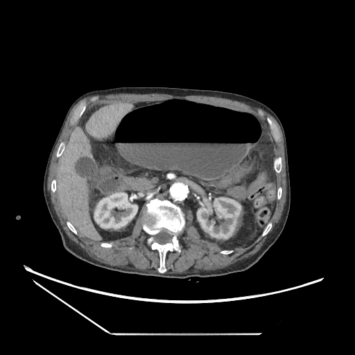
  • Plane projection: Axial
  • Aux modality: C+ arterial phase
  • Study findings: CT confirms the cecal volvulus with the lead point of the twist originating from the right lower quadrant, whilst the rest of the large bowel is largely empty. Associated small bowel distension and bowel wall thickening of the ascending and transverse colon. Other findings: status post NG tube insertion, an about 45 mm infrarenal, partially thrombosed abdominal aortic aneurysm, severe atherosclerosis, occlusion of the right common ilac artery, liver cysts, prostate enlargement, right inguinal hernia with small amount of free fluid, prior L3 vertebral compression.
  • Modality: CT
  • System: Gastrointestinal
  • Findings: Grossly distended large bowel loop with a 10 cm diameter and a large air-fluid level. The sigmoid contains a normal amount of air, and there are smaller air-fluid levels in the small bowel too. Findings altogether favor cecal volvulus with associated small bowel subileus.
Date Published: 9th May 2021
Source https://radiopaedia.org/cases/cecal-volvulus-9
Author Balint Botz
Permission
(Permission-reusing-text)
http://creativecommons.org/licenses/by-nc-sa/3.0/

Licensing:

Attribution-NonCommercial-ShareAlike 3.0 Unported (CC BY-NC-SA 3.0)

File history

Click on a date/time to view the file as it appeared at that time.

Date/TimeThumbnailDimensionsUserComment
current00:17, 13 July 2021Thumbnail for version as of 00:17, 13 July 2021512 × 512 (74 KB) (talk | contribs)Radiopaedia project rID:89212 (batch #6477-54 B53)

The following page uses this file:

Metadata