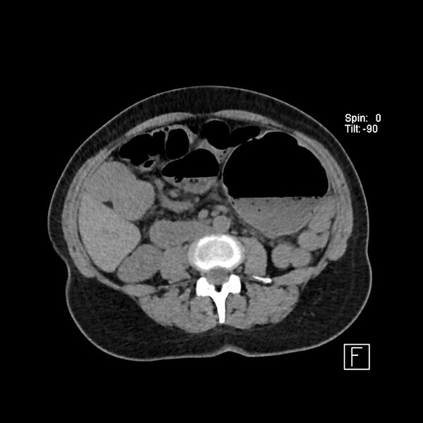
File:Cecal volvulus (Radiopaedia 90897-108397 Axial non-contrast 29).jpg
Jump to navigation
Jump to search

Size of this preview: 600 × 600 pixels. Other resolutions: 240 × 240 pixels | 480 × 480 pixels | 768 × 768 pixels | 1,024 × 1,024 pixels | 1,752 × 1,752 pixels.
Original file (1,752 × 1,752 pixels, file size: 247 KB, MIME type: image/jpeg)
Summary:
| Description |
|
| Date | Published: 3rd Jul 2021 |
| Source | https://radiopaedia.org/cases/cecal-volvulus-12 |
| Author | Antonio Rodrigues de Aguiar Neto |
| Permission (Permission-reusing-text) |
http://creativecommons.org/licenses/by-nc-sa/3.0/ |
Licensing:
Attribution-NonCommercial-ShareAlike 3.0 Unported (CC BY-NC-SA 3.0)
File history
Click on a date/time to view the file as it appeared at that time.
| Date/Time | Thumbnail | Dimensions | User | Comment | |
|---|---|---|---|---|---|
| current | 23:31, 12 July 2021 |  | 1,752 × 1,752 (247 KB) | Fæ (talk | contribs) | Radiopaedia project rID:90897 (batch #6475-29 A29) |
You cannot overwrite this file.
File usage
The following page uses this file: