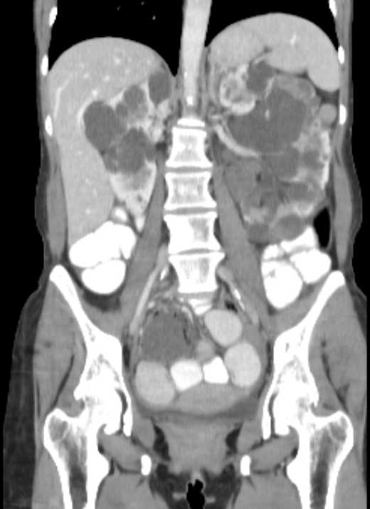
File:Cecal volvulus with pneumatosis coli (Radiopaedia 68572-78178 B 26).jpg
Jump to navigation
Jump to search
Size of this preview: 436 × 600 pixels. Other resolutions: 174 × 240 pixels | 520 × 715 pixels.
Original file (520 × 715 pixels, file size: 33 KB, MIME type: image/jpeg)
Summary:
| Description |
|
| Date | Published: 6th Jun 2019 |
| Source | https://radiopaedia.org/cases/cecal-volvulus-with-pneumatosis-coli |
| Author | Yair Glick |
| Permission (Permission-reusing-text) |
http://creativecommons.org/licenses/by-nc-sa/3.0/ |
Licensing:
Attribution-NonCommercial-ShareAlike 3.0 Unported (CC BY-NC-SA 3.0)
File history
Click on a date/time to view the file as it appeared at that time.
| Date/Time | Thumbnail | Dimensions | User | Comment | |
|---|---|---|---|---|---|
| current | 02:29, 13 July 2021 | | 520 × 715 (33 KB) | Fæ (talk | contribs) | Radiopaedia project rID:68572 (batch #6480-165 B26) |
You cannot overwrite this file.
File usage
The following page uses this file:
.jpg&oldid=742098)