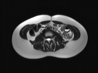
File:Cecoureterocele containing a calculus (Radiopaedia 80346-93729 C 28).jpg

From NC Commons
Jump to navigation Jump to search

Cecoureterocele_containing_a_calculus_(Radiopaedia_80346-93729_C_28).jpg(320 × 240 pixels, file size: 34 KB, MIME type: image/jpeg)

Summary:

Description
  • Radiopaedia case ID: 80346
  • Image ID: 53175295
  • Image stack position: 28/54
  • Plane projection: Axial
  • Aux modality: T2
  • Study description: MRI abdomen and pelvis
  • Study findings: MRI confirms a left-sided ureterocoele which descends through the urethra down into the vaginal introitus where it is manifest as a thin-walled blind ending cystic structure. This cystic structure contains a 7 mm calculus. The urinary bladder is partially filled and the contralateral right ureter is not well demonstrated.  The left ureter inserts slightly lower and more medially than expected, likely indicating an ectopic insertion close to the bladder neck.  The urinary bladder is partially collapsed but otherwise normal. The right distal ureter is normal in caliber and difficult to identify. The right kidney is normal in size and appearance.  The left kidney is slightly reduced in overall size with moderate dilatation of the renal pelvis up to 18 mm in AP diameter.  Mild fullness of the upper pole central calyces is present.  The lower pole calyces are essentially normal.  Only a single left ureter is readily identified. The vagina contains significant distension with fluid, perhaps representing vaginal secretions secondary to partial obstruction of the vaginal orifice by the cystic ureterocoele. Anteverted uterus is normal in size for age and demonstrates low dependent layering T2 hypointense fluid-fluid interface, likely representing hemorrhage from current menstruation.
  • Modality: MRI
  • System: Gynaecology
  • Findings: The palpable abnormality corresponds with a cystic structure anterior to the vagina and inferior to the symphysis pubis. A shadowing echogenic focus is present along its posterior aspect. There is an apparent left ureteric jet identified within this abnormality noted.  Definite direct communication between the abnormality and the bladder was not demonstrated.The right ureter was identified with a normal jet into the base of the bladder.  No left ureteric jet at the level of the bladder identified. Normal appearance of an anteverted uterus with volume of 33 ml.  Right and left ovaries have volumes of 3.2 ml and 1.7 ml respectively. Small amount of free fluid noted in the pouch of Douglas. Right and left kidneys measure at 108 mm and 80 mm respectively. No pelvicalyceal dilatation.
Date Published: 3rd Jan 2021
Source https://radiopaedia.org/cases/cecoureterocele-containing-a-calculus
Author James Harvey
Permission
(Permission-reusing-text)
http://creativecommons.org/licenses/by-nc-sa/3.0/

Licensing:

Attribution-NonCommercial-ShareAlike 3.0 Unported (CC BY-NC-SA 3.0)

File history

Click on a date/time to view the file as it appeared at that time.

Date/TimeThumbnailDimensionsUserComment
current02:57, 13 July 2021Thumbnail for version as of 02:57, 13 July 2021320 × 240 (34 KB) (talk | contribs)Radiopaedia project rID:80346 (batch #6481-79 C28)