File:Cecoureterocele containing a calculus (Radiopaedia 80346-93731 Axial non-contrast 41).jpg

From NC Commons
Jump to navigation Jump to search

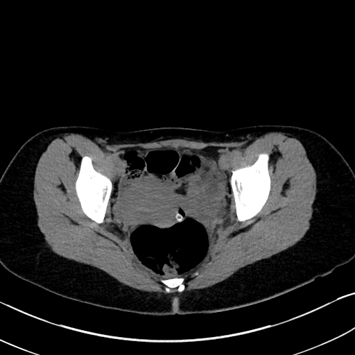
Cecoureterocele_containing_a_calculus_(Radiopaedia_80346-93731_Axial_non-contrast_41).jpg(512 × 512 pixels, file size: 89 KB, MIME type: image/jpeg)

Summary:

Description
  • Radiopaedia case ID: 80346
  • Image ID: 53175607
  • Image stack position: 41/74
  • Plane projection: Axial
  • Aux modality: non-contrast
  • Study description: CT cystogram
  • Study findings: Urethral catheter with balloon within the bladder and vaginal catheter with balloon within the upper vagina. Cystic structure with 7 mm calculus along its posterior aspect, is noted to the left of the bladder catheter balloon, consistent with known left ureterocele. Following urethral instillation of contrast into bladder lumen, the ureterocele appears as a filling defect outlined by contrast on all sides except its most posterior aspect, confirming its location within bladder. It is attached posteriorly to bladder wall very close to the bladder neck, suggesting ectopic inferomedial insertion of the left ureter. The ureter is difficult to identify given the distortion of anatomy from the vaginal cathether balloon. This study confirms the intravesical location of ectopic left ureterocele that had prolapsed through the bladder neck and urethra on the previous studies, and has now been reposed within the bladder lumen by the bladder catheter.
  • Modality: CT
  • System: Gynaecology
  • Findings: The palpable abnormality corresponds with a cystic structure anterior to the vagina and inferior to the symphysis pubis. A shadowing echogenic focus is present along its posterior aspect. There is an apparent left ureteric jet identified within this abnormality noted.  Definite direct communication between the abnormality and the bladder was not demonstrated.The right ureter was identified with a normal jet into the base of the bladder.  No left ureteric jet at the level of the bladder identified. Normal appearance of an anteverted uterus with volume of 33 ml.  Right and left ovaries have volumes of 3.2 ml and 1.7 ml respectively. Small amount of free fluid noted in the pouch of Douglas. Right and left kidneys measure at 108 mm and 80 mm respectively. No pelvicalyceal dilatation.
Date Published: 3rd Jan 2021
Source https://radiopaedia.org/cases/cecoureterocele-containing-a-calculus
Author James Harvey
Permission
(Permission-reusing-text)
http://creativecommons.org/licenses/by-nc-sa/3.0/

Licensing:

Attribution-NonCommercial-ShareAlike 3.0 Unported (CC BY-NC-SA 3.0)

File history

Click on a date/time to view the file as it appeared at that time.

Date/TimeThumbnailDimensionsUserComment
current03:16, 13 July 2021Thumbnail for version as of 03:16, 13 July 2021512 × 512 (89 KB) (talk | contribs)Radiopaedia project rID:80346 (batch #6481-41 A41)