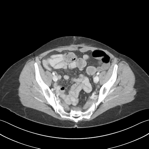
File:Cecum hernia through the foramen of Winslow (Radiopaedia 46634-51112 A 58).png
Jump to navigation
Jump to search
Cecum_hernia_through_the_foramen_of_Winslow_(Radiopaedia_46634-51112_A_58).png (512 × 512 pixels, file size: 72 KB, MIME type: image/png)
Summary:
| Description |
|
| Date | Published: 13th Jul 2016 |
| Source | https://radiopaedia.org/cases/caecum-hernia-through-the-foramen-of-winslow |
| Author | Bruno Di Muzio |
| Permission (Permission-reusing-text) |
http://creativecommons.org/licenses/by-nc-sa/3.0/ |
Licensing:
Attribution-NonCommercial-ShareAlike 3.0 Unported (CC BY-NC-SA 3.0)
File history
Click on a date/time to view the file as it appeared at that time.
| Date/Time | Thumbnail | Dimensions | User | Comment | |
|---|---|---|---|---|---|
| current | 04:06, 29 June 2021 |  | 512 × 512 (72 KB) | Fæ (talk | contribs) | Radiopaedia project rID:46634 (batch #5566-58 A58) |
You cannot overwrite this file.
File usage
The following page uses this file:
