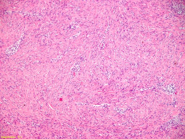
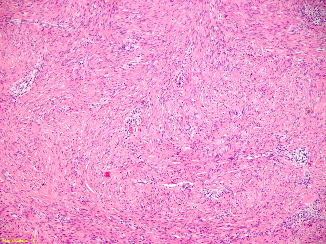
File:Cellular angiofibroma (vulva) - histology (Radiopaedia 43936-47432 40x H&E 1).jpg
Jump to navigation
Jump to search

Size of this preview: 800 × 600 pixels. Other resolutions: 320 × 240 pixels | 640 × 480 pixels | 1,024 × 768 pixels | 1,280 × 960 pixels | 2,560 × 1,920 pixels.
Original file (2,560 × 1,920 pixels, file size: 1.57 MB, MIME type: image/jpeg)
Summary:
| Description |
|
| Date | Published: 30th Mar 2016 |
| Source | https://radiopaedia.org/cases/cellular-angiofibroma-vulva-histology-1 |
| Author | Mikkaela McCormack |
| Permission (Permission-reusing-text) |
http://creativecommons.org/licenses/by-nc-sa/3.0/ |
Licensing:
Attribution-NonCommercial-ShareAlike 3.0 Unported (CC BY-NC-SA 3.0)
File history
Click on a date/time to view the file as it appeared at that time.
| Date/Time | Thumbnail | Dimensions | User | Comment | |
|---|---|---|---|---|---|
| current | 05:14, 13 July 2021 | | 2,560 × 1,920 (1.57 MB) | Fæ (talk | contribs) | Radiopaedia project rID:43936 (batch #6494-3 C1) |
You cannot overwrite this file.
File usage
The following page uses this file:
_-_histology_(Radiopaedia_43936-47432_40x_H%26E_1).jpg&oldid=1688181)