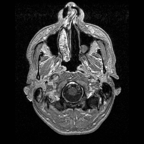
File:Central-variant posterior reversible encephalopathy syndrome (PRES) (Radiopaedia 43880-47358 Axial T1 C+ 1).jpg
Jump to navigation
Jump to search
No higher resolution available.
Central-variant_posterior_reversible_encephalopathy_syndrome_(PRES)_(Radiopaedia_43880-47358_Axial_T1_C+_1).jpg (288 × 288 pixels, file size: 54 KB, MIME type: image/jpeg)
Summary:
| Description |
|
| Date | Published: 1st Apr 2016 |
| Source | https://radiopaedia.org/cases/central-variant-posterior-reversible-encephalopathy-syndrome-pres |
| Author | Dr Sjoert Pegge |
| Permission (Permission-reusing-text) |
http://creativecommons.org/licenses/by-nc-sa/3.0/ |
Licensing:
Attribution-NonCommercial-ShareAlike 3.0 Unported (CC BY-NC-SA 3.0)
File history
Click on a date/time to view the file as it appeared at that time.
| Date/Time | Thumbnail | Dimensions | User | Comment | |
|---|---|---|---|---|---|
| current | 14:34, 14 July 2021 | | 288 × 288 (54 KB) | Fæ (talk | contribs) | Radiopaedia project rID:43880 (batch #6573-116 F1) |
You cannot overwrite this file.
File usage
The following page uses this file:
_(Radiopaedia_43880-47358_Axial_T1_C%2B_1).jpg&oldid=755312)