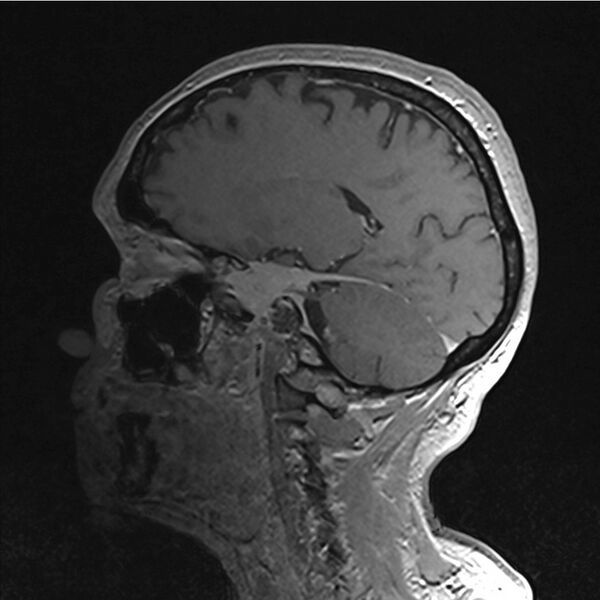
File:Central base of skull meningioma (Radiopaedia 53531-59549 Sagittal T1 C+ 43).jpg
Jump to navigation
Jump to search

Size of this preview: 600 × 600 pixels. Other resolutions: 240 × 240 pixels | 480 × 480 pixels | 953 × 953 pixels.
Original file (953 × 953 pixels, file size: 108 KB, MIME type: image/jpeg)
Summary:
| Description |
|
| Date | Published: 26th May 2017 |
| Source | https://radiopaedia.org/cases/central-base-of-skull-meningioma |
| Author | Dr Nikola Todorovic |
| Permission (Permission-reusing-text) |
http://creativecommons.org/licenses/by-nc-sa/3.0/ |
Licensing:
Attribution-NonCommercial-ShareAlike 3.0 Unported (CC BY-NC-SA 3.0)
File history
Click on a date/time to view the file as it appeared at that time.
| Date/Time | Thumbnail | Dimensions | User | Comment | |
|---|---|---|---|---|---|
| current | 07:47, 13 July 2021 |  | 953 × 953 (108 KB) | Fæ (talk | contribs) | Radiopaedia project rID:53531 (batch #6513-153 E43) |
You cannot overwrite this file.
File usage
The following page uses this file: