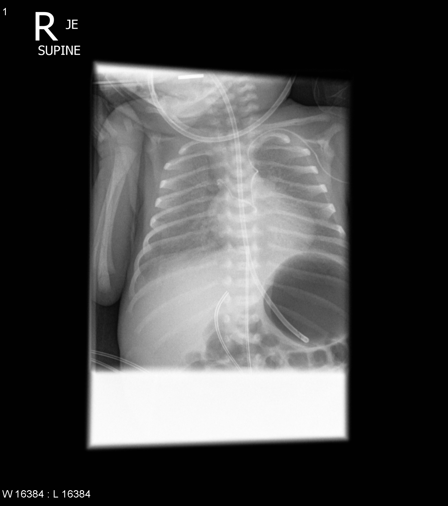
File:Central line in the accessory hemiazygos vein via the left superior intercostal vein (Radiopaedia 49913).jpg
Jump to navigation
Jump to search
Size of this preview: 532 × 600 pixels. Other resolutions: 213 × 240 pixels | 426 × 480 pixels | 681 × 768 pixels | 908 × 1,024 pixels | 1,534 × 1,730 pixels.
Original file (1,534 × 1,730 pixels, file size: 560 KB, MIME type: image/jpeg)
Summary:
| Description |
|
| Date | Published: 9th Dec 2016 |
| Source | https://radiopaedia.org/cases/central-line-in-the-accessory-hemiazygos-vein-via-the-left-superior-intercostal-vein |
| Author | Craig Hacking |
| Permission (Permission-reusing-text) |
http://creativecommons.org/licenses/by-nc-sa/3.0/ |
Licensing:
Attribution-NonCommercial-ShareAlike 3.0 Unported (CC BY-NC-SA 3.0)
File history
Click on a date/time to view the file as it appeared at that time.
| Date/Time | Thumbnail | Dimensions | User | Comment | |
|---|---|---|---|---|---|
| current | 11:05, 13 July 2021 | | 1,534 × 1,730 (560 KB) | Fæ (talk | contribs) | Radiopaedia project rID:49913 (batch #6524) |
You cannot overwrite this file.
File usage
The following file is a duplicate of this file (more details):
.jpg)
.jpg)
There are no pages that use this file.
.jpg&oldid=8853248)