File:Central line in the accessory hemiazygous vein via the left superior intercostal vein (Radiopaedia 49913).jpg
Jump to navigation
Jump to search

Size of this preview: 532 × 600 pixels. Other resolutions: 213 × 240 pixels | 426 × 480 pixels | 681 × 768 pixels | 908 × 1,024 pixels | 1,534 × 1,730 pixels.
Original file (1,534 × 1,730 pixels, file size: 560 KB, MIME type: image/jpeg)
Summary:
- Radiopaedia case ID: 49913
- Image ID: 26933544
- Modality: X-ray
- System: Vascular
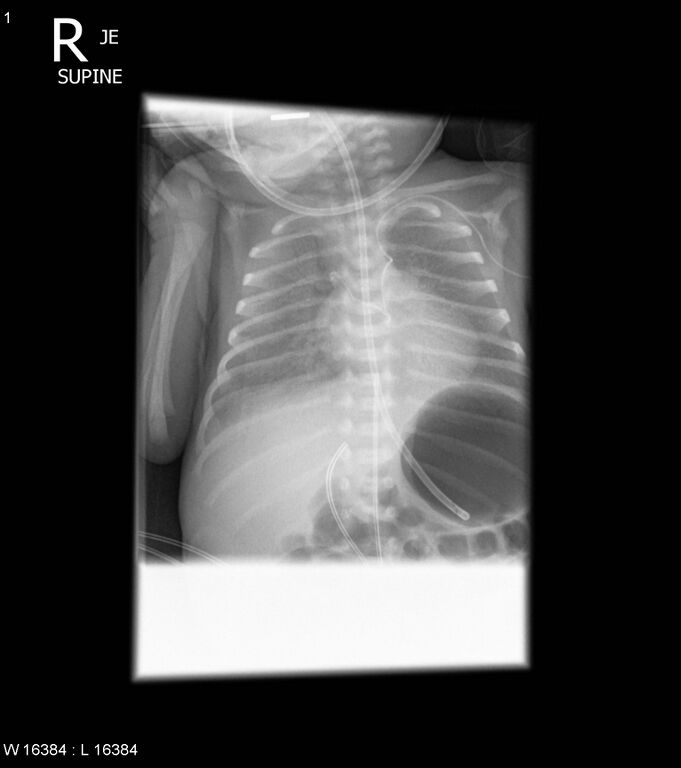
- Findings: A small bolus of iodinated contrast was injected through the newly inserted left subclavian CVL at the time of X-ray acquisition. The CVL passes from the left brachiocephalic vein into the left superior intercostal vein and then the accessory hemiazygous vein. Injected contrast streams accross the midline into the azygous vein and arch. NGT in stomach. UVC in ductus venosus. Stable granular opacification in the lungs in keeping with known HMD. Resolving medial right PTX.
- Published: 9th Dec 2016
- Source: https://radiopaedia.org/cases/central-line-in-the-accessory-hemiazygous-vein-via-the-left-superior-intercostal-vein
- Author: Craig Hacking
- Permission: http://creativecommons.org/licenses/by-nc-sa/3.0/
Licensing:
Attribution-NonCommercial-ShareAlike 3.0 Unported (CC BY-NC-SA 3.0)
File history
Click on a date/time to view the file as it appeared at that time.
| Date/Time | Thumbnail | Dimensions | User | Comment | |
|---|---|---|---|---|---|
| current | 12:44, 20 March 2021 | | 1,534 × 1,730 (560 KB) | Fæ (talk | contribs) | Radiopaedia project rID:49913 (batch #6292) |
You cannot overwrite this file.
File usage
The following file is a duplicate of this file (more details):
.jpg)
.jpg)
There are no pages that use this file.
.jpg&oldid=8859268)