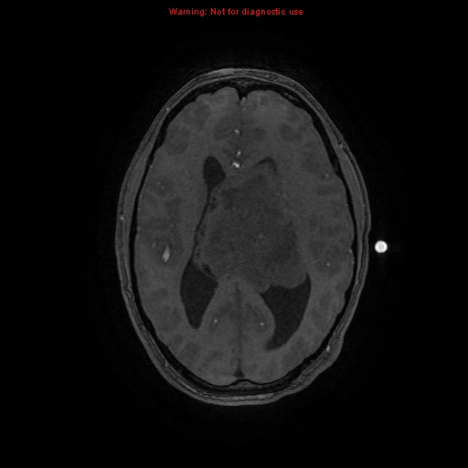
File:Central neurocytoma (Radiopaedia 13188-13206 Axial MRA 20).jpg
Jump to navigation
Jump to search
Central_neurocytoma_(Radiopaedia_13188-13206_Axial_MRA_20).jpg (512 × 512 pixels, file size: 65 KB, MIME type: image/jpeg)
Summary:
| Description |
|
| Date | Published: 11th Mar 2011 |
| Source | https://radiopaedia.org/cases/central-neurocytoma-7 |
| Author | Alexandra Stanislavsky |
| Permission (Permission-reusing-text) |
http://creativecommons.org/licenses/by-nc-sa/3.0/ |
Licensing:
Attribution-NonCommercial-ShareAlike 3.0 Unported (CC BY-NC-SA 3.0)
File history
Click on a date/time to view the file as it appeared at that time.
| Date/Time | Thumbnail | Dimensions | User | Comment | |
|---|---|---|---|---|---|
| current | 00:20, 14 July 2021 |  | 512 × 512 (65 KB) | Fæ (talk | contribs) | Radiopaedia project rID:13188 (batch #6538-73 H20) |
You cannot overwrite this file.
File usage
The following page uses this file:
