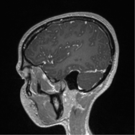
File:Central neurocytoma (Radiopaedia 37664-39557 Sagittal T1 C+ 23).png
Jump to navigation
Jump to search
Central_neurocytoma_(Radiopaedia_37664-39557_Sagittal_T1_C+_23).png (512 × 512 pixels, file size: 202 KB, MIME type: image/png)
Summary:
| Description |
|
| Date | Published: 18th Jun 2015 |
| Source | https://radiopaedia.org/cases/central-neurocytoma-16 |
| Author | RMH Neuropathology |
| Permission (Permission-reusing-text) |
http://creativecommons.org/licenses/by-nc-sa/3.0/ |
Licensing:
Attribution-NonCommercial-ShareAlike 3.0 Unported (CC BY-NC-SA 3.0)
File history
Click on a date/time to view the file as it appeared at that time.
| Date/Time | Thumbnail | Dimensions | User | Comment | |
|---|---|---|---|---|---|
| current | 13:26, 13 July 2021 |  | 512 × 512 (202 KB) | Fæ (talk | contribs) | Radiopaedia project rID:37664 (batch #6529-272 D23) |
You cannot overwrite this file.
File usage
The following page uses this file:
