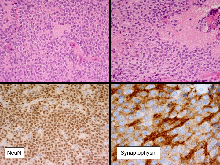
File:Central neurocytoma (Radiopaedia 37664-39585 A 1).png

From NC Commons
Jump to navigation Jump to search

Central_neurocytoma_(Radiopaedia_37664-39585_A_1).png(720 × 540 pixels, file size: 933 KB, MIME type: image/png)

Summary:

Description
  • Radiopaedia case ID: 37664
  • Image ID: 13811669
  • Study findings: MACROSCOPIC DESCRIPTION: 1. "Brain tissue": Fragments of soft gelatinous tissue up to 3mm. Portion for frozen section/smear. Remaining tissue for formalin plus glutaraldehyde (hold for EM). 2. "Brain tissue": Multiple pieces of soft tan tissue 17x15x3mm in aggregate. A1. MICROSCOPIC DESCRIPTION: 1&2. Paraffin sections show a moderately hypercellular tumor. Tumor cells have uniform medium sized round nuclei with granular chromatin and delicate processes and are arranged on lobules and solid sheets in a neuropil-like background. Scattered cells are undergoing apoptosis. No mitotic figures are identified. Small capillaries are prominent throughout the tumor but no multilayering of atypical cells around vessel lumena is seen. There is no necrosis. Immunohistochemistry shows that tumor cells are positive for synaptophysin and NeuN. GFAP and tyrosinase are negative. The features are of central neurocytoma (WHO Grade II). DIAGNOSIS: "Intraventricular tumor": Central neurocytoma (WHO Grade II).
  • Modality: Pathology
  • System: Central Nervous System
  • Findings: A large mass of heterogeneous signal and enhancement is seen within the frontal horn of the left lateral ventricle. The septum pellucidum is bowed to the left. The mass abuts the head of the caudate nucleus. There is obstructive hydrocephalus evident with transependymal spread on the right. Overall dimensions of the mass are 37.5 millimeters SI, 35 mm transverse and 36 mm AP. There is raised mild pulsatile and minimally increased choline within the mass. There is elevated cerebral blood volume (CBV) within the lesion. Conclusion: Findings compatible with a central neurocytoma.
Date Published: 18th Jun 2015
Source https://radiopaedia.org/cases/central-neurocytoma-16
Author RMH Neuropathology
Permission
(Permission-reusing-text)
http://creativecommons.org/licenses/by-nc-sa/3.0/

Licensing:

Attribution-NonCommercial-ShareAlike 3.0 Unported (CC BY-NC-SA 3.0)

File history

Click on a date/time to view the file as it appeared at that time.

Date/TimeThumbnailDimensionsUserComment
current14:46, 13 July 2021Thumbnail for version as of 14:46, 13 July 2021720 × 540 (933 KB) (talk | contribs)Radiopaedia project rID:37664 (batch #6529-1 A1)

There are no pages that use this file.