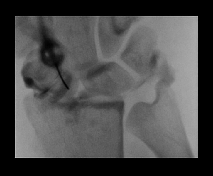
File:Central triangular fibrocartilage perforation (Radiopaedia 73346-84096 Dorsoventral 27).jpg
Jump to navigation
Jump to search

Size of this preview: 726 × 600 pixels. Other resolutions: 291 × 240 pixels | 581 × 480 pixels | 930 × 768 pixels | 1,240 × 1,024 pixels | 1,414 × 1,168 pixels.
Original file (1,414 × 1,168 pixels, file size: 139 KB, MIME type: image/jpeg)
Summary:
| Description |
|
| Date | Published: 8th Jan 2020 |
| Source | https://radiopaedia.org/cases/central-triangular-fibrocartilage-perforation |
| Author | Dai Roberts |
| Permission (Permission-reusing-text) |
http://creativecommons.org/licenses/by-nc-sa/3.0/ |
Licensing:
Attribution-NonCommercial-ShareAlike 3.0 Unported (CC BY-NC-SA 3.0)
File history
Click on a date/time to view the file as it appeared at that time.
| Date/Time | Thumbnail | Dimensions | User | Comment | |
|---|---|---|---|---|---|
| current | 14:11, 14 July 2021 | | 1,414 × 1,168 (139 KB) | Fæ (talk | contribs) | Radiopaedia project rID:73346 (batch #6572-27 A27) |
You cannot overwrite this file.
File usage
There are no pages that use this file.
.jpg&oldid=755170)