File:Centrifugal enhancement in a liver lesion (CEUS) (Radiopaedia 21641-21594 C 1).jpg
Jump to navigation
Jump to search

Size of this preview: 800 × 533 pixels. Other resolutions: 320 × 213 pixels | 640 × 426 pixels | 947 × 631 pixels.
Original file (947 × 631 pixels, file size: 154 KB, MIME type: image/jpeg)
Summary:
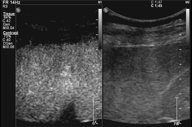
| Description |
|
| Date | Published: 6th Feb 2013 |
| Source | https://radiopaedia.org/cases/centrifugal-enhancement-in-a-liver-lesion-ceus |
| Author | Jan Frank Gerstenmaier |
| Permission (Permission-reusing-text) |
http://creativecommons.org/licenses/by-nc-sa/3.0/ |
Licensing:
Attribution-NonCommercial-ShareAlike 3.0 Unported (CC BY-NC-SA 3.0)
File history
Click on a date/time to view the file as it appeared at that time.
| Date/Time | Thumbnail | Dimensions | User | Comment | |
|---|---|---|---|---|---|
| current | 17:07, 14 July 2021 | | 947 × 631 (154 KB) | Fæ (talk | contribs) | Radiopaedia project rID:21641 (batch #6582-3 C1) |
You cannot overwrite this file.
File usage
There are no pages that use this file.
_(Radiopaedia_21641-21594_C_1).jpg&oldid=756258)