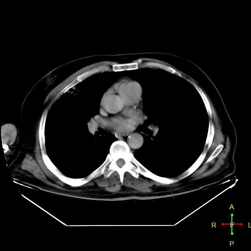
File:Centrilobular emphysema - with superimposed infection (Radiopaedia 24389-24683 Mediastinal 15).jpg
Jump to navigation
Jump to search
Centrilobular_emphysema_-_with_superimposed_infection_(Radiopaedia_24389-24683_Mediastinal_15).jpg (512 × 512 pixels, file size: 24 KB, MIME type: image/jpeg)
Summary:
| Description |
|
| Date | Published: 11th Aug 2013 |
| Source | https://radiopaedia.org/cases/centrilobular-emphysema-with-superimposed-infection |
| Author | Ahmed Abdrabou |
| Permission (Permission-reusing-text) |
http://creativecommons.org/licenses/by-nc-sa/3.0/ |
Licensing:
Attribution-NonCommercial-ShareAlike 3.0 Unported (CC BY-NC-SA 3.0)
File history
Click on a date/time to view the file as it appeared at that time.
| Date/Time | Thumbnail | Dimensions | User | Comment | |
|---|---|---|---|---|---|
| current | 17:26, 14 July 2021 |  | 512 × 512 (24 KB) | Fæ (talk | contribs) | Radiopaedia project rID:24389 (batch #6587-43 C15) |
You cannot overwrite this file.
File usage
There are no pages that use this file.
